Материал: BASIC CLINICAL SYNDROMES IN INTERNAL DESEASES CLINIC
6. The syndrome of accumulation of air in the pleural cavity
REASONS: occurs when bronchi communicate with the pleural cavity (subpleural location of the tuberculous cavity, abscess), with chest injury, spontaneous, artificial pneumothorax, etc. SYMPTOMS:
respiratory dyspnea;
On examination - protrusion and limitation of mobility of the corresponding side, smoothing of intercostal spaces;
If the air pressure in the pleural cavity is large, there is an increased resistance to intercostal spaces;
Voice jitter is weakened or absent;
With percussion: on the sore side - a loud tympanic tone, sometimes with a metallic tint. If the air pressure is high, the percussion tympanic tone may be blunted.
Auscultatory: breathing and bronchophony are weakened or absent (closed pneumothorax). If there is a message between the pleural cavity and the bronchus, amphoric breathing can be heard, bronchophony in this case is enhanced;
11
X-ray: a bright pulmonary field without pulmonary pattern, and closer to the root - the shadow of a compressed lung. With intense pneumothorax - displacement of the mediastinal organs to the healthy side;
With the simultaneous accumulation of liquid and air (Hydropneumothorax) in the pleural cavity, the horizontal upper limit of dullness corresponds to the level of the liquid. Dullness easily changes its boundary when changing the position of the patient's body. This border usually remains horizontal, with a loud tympanic sound above it. When succussion is determined by the noise of the splash (taking Hippocrates) in the chest.
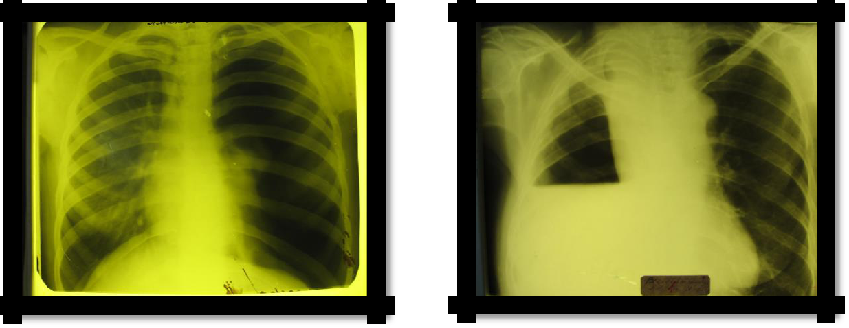
Pic 2.6 Pneumothorax Pic 2.7 Hydropneumothorax
7. Bronchiectasis Syndrome
REASONS: clinically manifests itself in the development of a chronic purulent process in altered
dilated bronchi.
SYMPTOMS:
Cough with sputum (mucopurulent, sometimes three-layer), mainly in the morning (2/3 of daily sputum: "morning toilet of the bronchi"), hemoptysis. "Dry" bronchiectasis has no clinical manifestations, with the exception of hemoptysis;
Hard breathing, dry and wet rales are possible with a compact arrangement of bronchiectasis;
X-ray: amplification, ―cellularity‖ of the pulmonary pattern (not always detected). The "gold standard" of the study is bronchography (contrasting of the bronchi with iodolipol), reveals the characteristic saccular and spherical extensions of the bronchi. Recently, computed tomography
(CT) with contrast has been widely used.
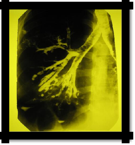
Pic 2.8 Bronchography
12
8. Syndrome of lung atelectasis
Usually distinguished: obstructive, compression and distensional (functional) atelectases
I. REASONS: obstructive atelectasis (bronchus is blocked by a tumor, thick mucus or a foreign body):
SYMPTOMS:
chest lag in the act of breathing;
voice jitter is not detected;
blunting or tympanic percussion sound;
respiration and bronchophony are not determined;
X-ray: intercostal space narrowed, homogeneous darkening in the atelectasis and mixing of the mediastinum to the affected side (Goltsknecht-Jacobson symptom), high standing of the diaphragm.
II. REASONS: compression atelectasis (the bronchus is free, the lung is squeezed by exudate, air, a large spleen through the diaphragm, etc.).
SYMPTOMS in the area of the dormant lung:
voice trembling enhanced;
blunt-tympanic percussion sound;
Weakened vesicular breathing, often with a bronchial tinge, crepitus is possible. Bronchophony is enhanced;
X-ray: increased pulmonary pattern or homogeneous darkening in the area of atelectasis.
The clinical picture of the disease that caused atelectasis (see syndromes of accumulation of air and fluid in the pleural cavity).
III. REASONS for distensible atelectasis (weakness of the respiratory movements, decreased tone of the respiratory muscles, when the pressure from the organs of the abdominal cavity, the lungs do not completely straighten and contract - tightening).
SYMPTOMS: the lower border of the lungs is 1-2 ribs above the norm, above the lower sections breathing is weakened by vesicular, crepitus and ―rattling‖ rales, which often disappear after several deep breaths.
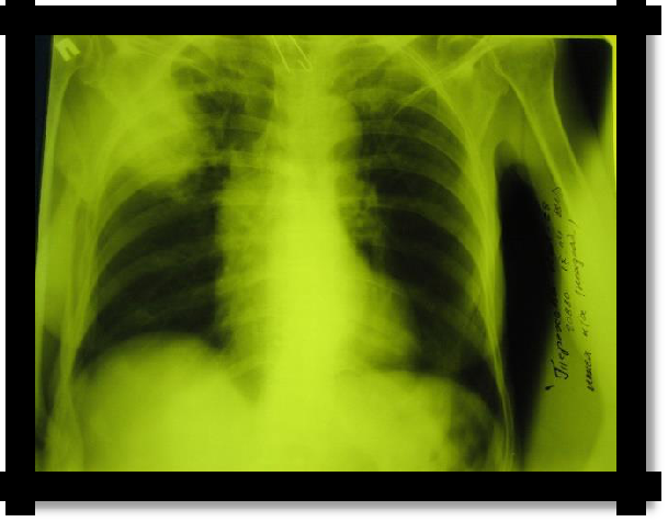
Pic 2.9 Obstructive atelectasis
13
9. Syndrome of dry pleurisy (pleuritis)
REASONS: more often a consequence of other diseases (pneumonia, tuberculosis, systemic
diseases of the connective tissue, uremia, etc.).
SYMPTOMS:
Pain in the chest, which intensifies with deep breathing and coughing;
Superficial breathing, limiting the mobility of one half of the chest. The patient lies on the affected side;
Percussion: limitation of the mobility of the lower border of the lung on the side of the lesion;
The noise of pleural friction during auscultation, sometimes confirmed by palpation;
With diaphragmatic pleurisy, pain radiates to the upper half of the abdomen or along the phrenic nerve to the neck. With apical pleurisy - pain during palpation of the trapezius muscles and pectoralis major muscles (Sternberg and Pottenger symptoms, respectively).
X-ray: on the side of the lesion, a high standing of the diaphragm, limitation of excursion of the lower edge with fluoroscopy, signs of a disease that caused pleurisy.
10. Respiratory failure syndrome
REASONS: is a complication of any severe pathological process or disease or a secondary lesion of the respiratory system.
Respiratory failure is a pathological condition of the body in which the normal gas composition of the blood is not maintained or is achieved due to the strain of compensatory-adaptive mechanisms leading to a decrease in the functional capabilities of the body. SYMPTOMS:
The most constant and leading sign of respiratory failure is shortness of breath. The severity of dyspnea reflects the degree of respiratory failure.
According to the classification of A.K. Dembo, proposed back in 1954 and currently used, 3 degrees of respiratory failure are distinguished:
degree. Latent (asymptomatic) RF (shortness of breath with moderate and significant physical exertion, the gas composition of the blood at rest is not changed).
degree. Compensated RF (shortness of breath with slight physical exertion, at rest - hypoxemia, no hypercapnia, compensatory mechanisms are already at rest).
degree. Decompensated RF (shortness of breath at rest, hypoxemia and hypercapnia).
In the diagnosis of respiratory failure, Stange tests are widely used - breath holding at the height of inspiration and Gencha - holding the breath at the height of exhalation. The lower limits of the norms of these samples are 40 and 20 s.
Another mandatory and universal sign of respiratory failure is diffuse cyanosis.
With severe respiratory failure are possible: forced body position (for example, the position of orthopnea in an attack of bronchial asthma), erythrocytosis, tachycardia, a change in the gas composition of the blood (pulse oximetry).
Change in spirometric indicators.
obstructive;
restrictive;
mixed.
Table 2.2 Classification of respiratory failure by the state of blood gases
-
degree
Р О2, мм рт.ст.
Sp02, %
Normal level
>80
>95
I
60-79
90-94
II
40-59
75-89
III
<40
<75
11. Pulmonary heart failure (pulmonary heart)
It is divided into acute and chronic
Reflects the dependence of the development of right ventricular cardiac decompensation (pulmonary heart) in severe diseases of the bronchopulmonary system (Euler-Lillestrand reflex). Chronic
Stage 1 - purely pulmonary (respiratory failure is observed, pulmonary hypertension is formed) Stage 2 - pulmonary + compensated right ventricular heart failure (right ventricular hypertrophy develops against pulmonary hypertension)
Stage 3 - pulmonary + decompensated right ventricular heart failure (congestion in a large circle
of blood circulation).
SYMPTOMS:
Expansion of the boundaries of the relative dullness of the heart to the right
accent 2 tones over the pulmonary trunk
Signs of congestive right ventricular heart failure
pulmonary hypertension according to ultrasound
12. Adult respiratory distress syndrome
Adult respiratory distress syndrome (RDSV) is an acute respiratory failure that occurs with acute lung injuries of various etiologies, with the obligatory development of non-cardiogenic pulmonary edema (interstitial, then alveolar). A synonym is shock lung.
Etiology: sepsis, shock, chest injuries, aspiration of water or stomach contents, pneumonia, inhalation of irritating and toxic substances, prolonged exposure to high altitude, venous fluid overload, severe metabolic disturbances, massive blood transfusions, autoimmune diseases.
RDSV develops after a latent period - a period of exposure to the etiological factor (about 24 hours). Etiological factors lead to the accumulation of active white blood cells and platelets in the pulmonary capillaries and interstitial lung tissue, the release of biologically active substances.
The acute phase. There is pulmonary edema, hypoventilation, microatelectases, impaired diffusion of oxygen and carbon dioxide. It ends with recovery or transition to the subacute phase. The subacute phase is characterized by the presence of interstitial and bronchoalveolar inflammation.
The chronic phase is characterized by the development of interstitial fibrosis (already from the 2nd week of the disease).
The clinical picture.
Acute respiratory failure: severe shortness of breath, diffuse cyanosis, participation of auxiliary muscles in the act of breathing, tachycardia, hypoxia, hypercapnia.
16
2. Lowering blood pressure.
3.Symptoms of interstitial pulmonary edema: decreased elasticity of the chest, dullness of percussion sound, more in the posterior - lower parts of the chest, hard breathing, there may be dry wheezing.
With the appearance of alveolar pulmonary edema - moist rales, crepitus, cough with foamy pink sputum.
Syndrome of acute pulmonary heart and pulmonary hypertension (pressure in the pulmonary artery rises (> 30/15 mm Hg), but unlike cardiogenic pulmonary edema, the normal pressure is pulmonary wedge jamming (<15 mm)).
Multiple organ failure. Metabolic acidosis (pH <7.3).
X-ray - pronounced interstitial pulmonary edema, bilateral shadows of irregular cloud shape.
2.1. TEST TASKS
(Choose one or more correct answers)
Indicate the most characteristic changes in the chest with the syndrome of fluid accumulation in the pleural cavity:
a decrease in half of the chest, its retraction and lag during breathing
an increase in half the chest, smoothing of the intercostal spaces, chest lag during breathing
the chest is not changed, the lag of half of the chest during breathing