Материал: BASIC_SURGICAL_TECHNIQUES budapest
C
A
B
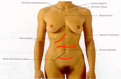
Figure 68. Transverse and oblique laparotomies
A. Upper transverse laparotomy, B. Lower transverse laparotomy, C. Paracostal laparotomy
Muscle-splitting incisions
In these types of incisions the fibers of the abdominal wall muscles are not cut but separated from each other alongside their courses. Advantage: the possibilty for developments of postoperative hernia is rare. Disadvantage: it gives a limited exposure and is helpful only in the case of a sure diagnosis.
Lower median muscle-splitting incision (Pfannenstie incision)
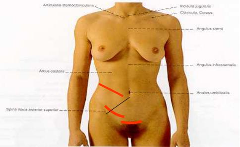
A transverse incision about 2-3 finger breadths above the symphysis pubis and between the two external edges of the rectus muscles. The anterior rectus sheaths are cut and the rectus muscles are retracted bluntly. It is used mainly in OBG (Figure 69. A).
Upper lateral muscle-spliting incision
It is used exclusively in the newborn babies for the purpose of pyloromyotomy.Performed on the right side (Figure 69. B).
Lower lateral muscle-splitting incision ((McBurney incision)
One of the most common types of incisions they are. A 4-6 cm long incision made at the middle and outer 1/3 of an imaginary line which connects the right ant. sup. iliac spine to the umbilicus. 1/3 of this incision is located above and 2/3 of it below this imaginary line. Starting with separation of the fibers of the ext. oblique muscle we finally explore the appendix (Figure 69. C).
71
B
C
A
Figure 69. Muscle-splitting incisions
A. Pfannenstiel incision, B. Upper lateral muscle-splitting incision, C. McBurney incision
6.3.2. Laparotomy on the posterior abdominal wall
Oblique posterior approach (Bergmann-Israel incision)
Starting from the lower egde of the 12 th rib we go towards the ant. sup. iliac spine and then parallel with the Poupart lig. we end our incision at the ant. abdominal wall. To have a better exposure, it is also necessary to resect the12th rib subperiosteally.
Vertical posterior approach
It is a rarely used incision which is made alongside the external edge of the deep back muscles.
6.3.3. Thoracolaparotomy
In the csae of big tumors of the liver, tumors of the kidney, possibly the total gastrectomy, operations done around the cardia region, and oesophageal tumors, when there is a need to open the thoracic and the abdominal cavities simultaneously, we apply this incision. It is an upper transrectal or an upper transverse laparotomy whieh is running alongside the 7th intercostal space.
6.3.4. Skin incisions and the positions of the trocars in laparoscopic surgeries
Within the last 2 decades, the most significant development in surgery was the appearance of the laparoscopic surgery. During the conventional open surgery the surgeon has the possibility to palpate the abdominal cavity and exclude the presence of other diseases. Disadvantages of this type of approach are: long hospital stay and rehabilitation period, returning to work after a long period of time, decreased ability to perform the customary work, and possibilty for the postoperative hernia.
In laparoscopic approach we can inspect the abdominal cavity only visually. The degree of postoperative pain and the duration of hospitalization are significantly decreased. The recovery is faster and patient can go to work even a few days after operation. The most commonly preformed laparoscopic surgery is the laparoscopic cholecystectomy. The Figure 70. shows the position of the trocars.
72
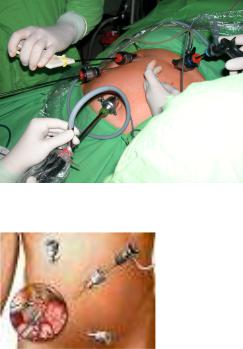
The right lower abdominal cavity can also be approached by laparoscopic instruments which is primarily important from standpoint of differential diagnoses. Nowdays, more and more appendectomies are done by this method which consequence is abscence of usual appendectomy scar (Figure 71.). In the case of recurrent lower abdominal pain this can cause a dilemma for uncareful practioners.
The minilaparotomy incisions needed in hand-assisted laparoscopic surgeries are more frequently located at the midline.
Figure 70. Trocars placed during a laparoscopic cholecystectomy
Figure 71. Trocars placed during a laparoscopic appendectomy
7. BASICS OF THE LAPAROSCOPIC SURGERY
Nowadays, the reduction of the surgical load is characterstic in the surgical interventions. The laparoscopic technique which basically changed the classic surgical mentality, was the milestone in this aspect of thinking. It was certified that the extent of the surgical incision plays an important role in the patient’s recovery.The multidisciplinary approach replaced the virtuoso surgeon -who could solve everything aloneand certified that the technique is able to improve the surgeon’s skill and his operative possibilities.
In 1902 Kelling called the method as laparoscopy. A word of Greek origin which means: the inspection of the soft tissue (αραπαλ: soft tissue, κσcοωεπ: inspect).
73
The technical developmets, training operations and the patients’ increased demands for the minimally invasive surgeries contributed to the wide spread of the laparoscopic surgeries
7.1. History
1901: D.O. Ott (Saint Petersburg) gynecologist – ventroscopy: through a colposcopic orifice: head mirror + speculum
1901: G. Kelling (Drezda) – coelioscopy with a cystoscope and in an dog, pneumoperitoneum for the first time
1910: H.C. Jacobeus (Stockholm) – laparoscopy for the first time in human 1929: H. Kalk -135 0 forward viewing optic (i.e.lens system): air between the lens 1933: C. Fervers – laparoscopic adhesiolysis
1938: János Veres (Kapuvár) – pulmonologist – treatment of the pneumothorax special needle to create the pneumoperitoneum
1960: K. Semm (gynecologist) - automatic insufflator, appendectomy
1966: H.H. Hopkins – optical system: instead of air, glass tubes are between the lens (loss of light ↓)
1985: E. Mühe: first cholecystectomy ”keyhole surgery” – lecture without any response 1987: P. Mouret - laparoscopic cholecystectomy. Although he did not published anything
about it. However, they consider him as the person who performed the first laparoscopic
cholecystectomy
1990: first laparoscopic cholecystectomy in Hungary – Pécs, Tibor Kiss
7.2. Comparison of the open and laparoscopic techniques Disadvantages of open surgery
•big exposure , more operative trauma
•the postoperative pain depends mostly on the size of the surgical wound
•it is harmful to keep the body cavity open for a long time (vaporization, drying, etc.)
•danger of secondary injuries during exposure (i.e. intestine, spleen, lung)
•the increased possibility for later adhesions
•the bigger the wound is, the higher the possibilty for postoperative complications (i.e. infection, hernia, etc.) is
Advantages of the laparoscopic technique
•smaller exposure, less operative trauma
•less postoperative pain
•reduced numbers of wound infection and hernia
•less postoperative adhesions
•shorter postoperative healing period and duration of the hospitalization
•cosmetic adventages
7.3. Instruments and steps of the laparoscopic surgeries
7.3.1. Creation of a pneumoperitoneum
The space needed in the abdominal cavity to perform the laparoscopic surgeries is produced by insufflating it with a gas.This is called the generation of the pneumoperitoneum. At the begining it was done by pumping the air into the abdominal cavity. Nowdays, CO2 due to the following characteristics is considered to be a safe gas:
-it is absorbed rapidly by the peritoneum,
74
-it is dissolvable in the blood,
-it is excreted rapidly from the body, and
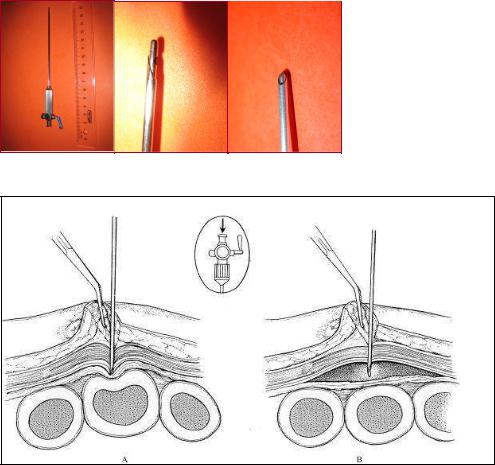
-it can be used simultanously with the electrocautery device with no danger of the explosion. Disadvantage: The chance for development of cardiac arrhythmias is more in the case of application of CO2. Due to the danger of the hypercarbia, it is very important to monitorize the patient.To create a pneumoperitoneum, we use the Veres needle (Figure 72.).
Figure 72. Veress needle
Figure 73. Insertion of the Veress needle into the abdominal cavity
The internal part of the needle, which is a blunt obturator, retracts on contact with abdominal wall to reveal a cutting tip to enterwithout any difficulty -into the peritoneal cavity. Following removal of the resistance, the blunt obtruator comes out again to prevent injury to the abdominal visceras (Figure 73.).
The insufflator is maintaining the pneumoperitoneum. From standpoint of the safety of the surgery, it is necessary to monitorize the intraabdominal pressure, the flow rate of the gas, and the volume of the used gas continuously. To avoid the over-insufflation, which can hinder the venous return, the intraabdominal pressure should be less than 15-20 mmHg. The safety system of the modern insufflator prevents the pressure exceeding the set limit (e.g. 15 mmHg) (Figure 74.).
75