Материал: BASIC_SURGICAL_TECHNIQUES budapest
-altered anatomical relations
-atypical symptoms
-possibilty for foetal diseases
Menstration
In the past, there was a tendency to avoid surgery on a menstrating woman due to increased psychic unstability, possible increase in bleeding tendency, and increased hygienic demands seen during this period. Nowdays, the menstration is not considered as an obstacle to do the operation.
Overfeeding as a factor increasing the surgical risk
-respiratory disturbancy(usually restrictive): deteriorating the gas exchange, increased respiratory function
-decresed cardiac reservoirs
-difficulty with intubation (regurgitation)
-disturbancies with wound healing
-thromboembolism
Immunological factors
Immunosuppresion (transplanted patient), use of cytostatics (tumorous patient), AIDS and so on increased possibilty for infection, frequent wound healing disturbancies.
Oncological patients’ own problems
-chemotherapeutic agents
-radiotherapy (local inflammation)
-decreased function of the immune system
-paraneoplastic syndromes e.g. deep venous thrombosis
Increased age as a factor influencing the surgical risk-elderly patient
-elderly patient: age > 65 years old (the biological age is important and not the calender age)
-to assess the expected benefits, risks, and the patient’s survival
-to estimate the interactions between the drugs, used by patient for a long period of time and the necessary drugs which are given during the perioperative period
-cardiopulmonary deficiency is the cause of the death in most cases
Estimation of the surgical risks
It means to examine the followings:
-cardiovascular state,
-respiratory system,
-metabolic state,
-renal function,
-liver function,
-endocrine balance,
-homeostasis,
-immune system.
Examinations
-physical examination
-laboratory examination
-radiological examinations (US, CT, MRI, isotop, DSA, and so on)
-instrumental examination (endoscopy, biopsy, cytological examinations)
66
Heart and circulation:
-pulse, blood pressure
-ECG
-Echocardiography
-coronarography
-isotop
Increased cadiac risks:
-aortic stenosis, mitral stenosis
-dysrhythmia
-AMI: within 3 weeks mortality: 25%
-AMI: within 6 weeks mortality: 5%
-DM silent ischemia 25%
Lung, breathing:
-Chest X-ray
-Respiratory function
-Blood gas analysis (preparation for the surgery: respiratory physiotherapy and inspiratory treatment)
Laboratory examinations:
-blood count
-blood group
-bleeding and clotting times
-liver function tests
-renal function tests
-examination of the metabolic processes
-fluid and electrolyte balance
-plasma protein level
Diet regulations
If it is possible, you should prevent the deterioration of the nutritional status of the patient during the processes of the preoperative examination and preparation. Sometimes, the nutritional therapy is a part of preoperative preparation. To have a safe general anesthesia, the patient should avoid eating (fasting 6 hours prior to surgery).
Preoperative nutritional therapy
First, you should consider the natural oral feeding. If it is not possible, then the nasogastric, duodenal or jejunal tubes are the most appropriate ways of feeding. The parentral feeding is done through the peripheral or central venous catheters. The burned, tumorous, polytraumatized, and septic patients need the highst amount of energy.
Slag deprivation
-Diet: liquids for 2-3 days or a low-residue diet
-Enema: In the case of major abdominal surgeries (or those operations which involve the intestinal system), there is a need to make the intestinal tract empty.
-to make the stomach empty: In the case of pyloric stenosis, the nasogastric tube can remove the gastric contents and also lead to the gastric lavage(antibiotic).
Urinary catheter
67
It is needed in the case of long-lasted operations which are running with loss of a large amount of fluids.
Thrombosis prophylaxis
-Drugs:
-Heparin derivatives: Na-heparin, Ca-heparin, low molecular weight heparins
-Platelet aggregation inhibitors (e.g. Aspirin and Colfarit)
-Coumarin derivatives (e.g. Syncumar)
-Physical:
-early mobilization
-compression (elastic bandages)
-bed-side bicycle
-keeping the lower extremities at a high level
Psychic preparartion
That is natural for the patient to fear of the operation and its unwanted consequences. The surgeon should deals with the patient’s psychic state. He/she should carefully evaluate the indications and contraindications and choose the best possible intevention.
Legal aspects of the operations
-informing the patient (tumorous patients!)
-patient’s written consent (this should also include those decisions which may be made by the surgeon intraoperatively).
-in the case of children, the parents or the legal representative should give the informed written consent.
6.3 Surgical approaches
6.3.1. Laparotomy on the anterior abdominal wall
The direction of the incision can be: verical, transverse, or oblique. Vertical incisions:
-upper, lower, middle, or total median laparotomy
-paramedian laparotomy
-vertical transrectal laparotomy
-pararectal laparotomy
Transverse incisions:
-horizontal transrectal laparotomy
-Pfannensteil incision
Oblique incisions:
-McBurney-incision
-inguinal transmuscular laparotomy
-paracostal laparotomy (Kocher incision)
-subcostal laparotomy
Vertical incisions
Upper median laparotomy
The incision is made from xyphoid process to the umbilicus. Advantages: insures a quick and wide exposure, quickly and easily can be elongated and closed. Disadvantages: the fibrous tissue of the white line (linea alba) is cut and the the sutures are upon tensions at both sides
68
which can be a cause for a later postoperative hernia (Figure 67. A).
Lower median laparotomy
The incision is made from umbilicus to symphysis pubis. The advantages and disadvantages are the same as those for an upper median laparotomy. About 2/3 of sterile wound disruption happens folowing such a this incision (Figure 67. B).
Middle median laparotomy
An 8-10 cm long incision. Half of it is located above and the other half below the umbilicus. At the umbilcus the incision is curved towards the left side. Advantage: from a small incision we can inspect both the upper and the lower part of the abdominal cavity.
Total median laparotomy
The incision is made from xyphoid process to the syphysis pubis. It gives an excellent exposure but injures the statistic of the abdominal wall significantly. The patient is predisposed to the postoperative wound disruption. It also makes the postoperative coughing difficult, increases the danger of pneumonia, and can cause constipation. This incision is generally used in the case of extended abdominal operations (Figure 67. A+B).
Paramedian laparotomy
It is generally used only above the umbilicus. About 2 cm right (an parallel) to the midline cut the skin, subcutaneous tissue and the anterior leaflet of the rectus sheath. The rectus muscle is retracted. Following this, the posterior rectus sheath is also cut. The later scar will be strong and the possibilty for development of a henia is rare (Figure 67. C).
Transrectal laparotomy
About 2-3 cm right to the midline cut the skin, subcutaneous tissue, and the anterior leaflet of the rectus sheath. Then, separate the fibers of the rectus muscle from each other bluntly to be able to cut the posterior leaflet of the rectus sheath (together with transversalis fascia and the parietal peritoneum)(Figure 67. D).
Pararectal laparotomy
A vertical incision is parallel to the rectus muscle. Due to the denervation of the muscles the abdominal wall becomes significanly weakened. It is danger for development of a huge hernia. It is not advisable (Figure 67. E).
Lateral transmuscular laparotomy
The incision is made starting from a point located 2-3 cm lateral to the external edge of the rectus muscle. The longest incision of such starts at the lower edge of the 10 th rib and runs till the level of the anterior sup. ilac spine. The” pararectal” and ”an incision made alongside the semilunar line of Spiegel” are not the ideal incisions because they weaken the abdominal wall significantly. They are not advisable (Figure 67. F).
69
D C A
E
F
B
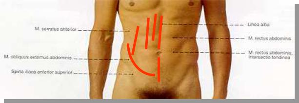
Figure 67. Vertical laparotomies
A.Upper median laparotomy, B. Lower median laparotomy, A+B Total median laparotomy, C. Paramedian laparotomy, D. Transrectal laparotomy, E. Pararectal laparotomy, F. lateral transmuscular laparotomy
Transverse and oblique laparotomies
They cause less injury to the nerves of the abdominal wall muscles. In this way, the possibilties for postoperative sterile wound disruption and later hernia are less.
Upper transverse laparotomy
The incision is made at the area beween the xyphoid proc. and the umbilicus, starting from one external edge of the rectus muscle and ending at its other edge (It is at the border line beween the middle and lower 2/3) (Figure 68. A). This incision can be made larger by elongating it at both of its lateral sides (even up to the middle axillary lines). This incision rarely injures the abdominal wall muscles. The innervations of these muscles are not injured and the wound heals with development of a strong scar. In upper abdominal surgeries we can combine the upper median incision with a transverse incision. Such this incision is called: the Mercedes-Benz incision.
Lower transverse laparotomy
This a mild concave incision a few centimeter below the umbilicus (Figure 68. B). One or both rectus muscles are cut. Sometimes, we only cut the rectus sheaths and do not incise the muscle itself. The incision can be elongated laterally.
Paracostal laparotomy
A curved incision started from xyphoid proc. to the lateral side of the abdominal wall. It is located 2-3 cm below and parallel with the costal margins (Figure 68. C). The possibilty for development of a postoperative hernia is high. On the left side, this incision is helpful in performing a splenectomy. On right side it can be used to perform an open cholecystectomy. Nowdays, with application of the laparoscopic cholecystectomy we can avoid the postoperative pain and complications of such this incision!
70