Материал: BASIC_SURGICAL_TECHNIQUES budapest
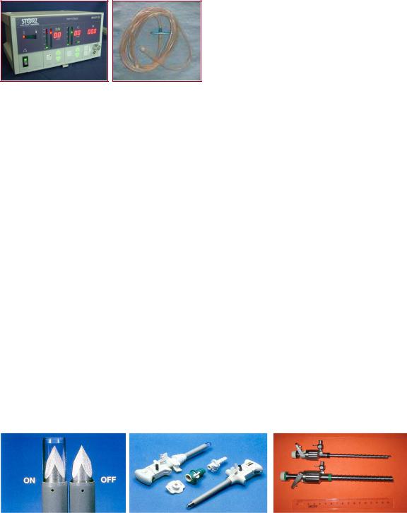
Figure 74. Insufflator and its connecting gas tube
7.3.2. Entry into the abdominal cavity
Once the pneumoperitoneum is established, a “trocar-port” assembly must be inserted to allow the passage of the laparoscope and operating instruments into the peritoneal cavity. The main parts of a trocar-port assembly are: spit (or obturator, or trocar), cannula (or port), and the valve.
The length, thickness, and the shape of the tip of the trocar can be different. The automatic trocar-port is supplied with a safety shield that reduces injury to organs during insertion: it has a plastic safety shield that retract to expose the sharp tip during the insertion, and spring back upon entry into the peritoneal cavity. In the most up-to-date trocar-port the trocar itself spring back after entring into the peritoneal cavity.
The port (cannula) is furnished with a valve.It makes possible to insert the optic and different working instuments into the abdominal cavity. The tissue parts can also be removed through the port. The external diameter and the length of it can be between 5 to 25 mm and 11.5 to 17 cm respectively. The valve, which is a springy metal inset lying perpendicular to the axis of the port, prevents the gas to escape from the abdominal cavity. The automatic socalled „”tilting valve” is opened by advancing the instrument in the port and is automatically closed after removal of the instrument (Figure 75.).
There are many holes at the distal part of the port. These let the gas enter into the abdominal cavity without needing to reach to the distal lens of the optic, which otherwise will lead to the disturbing condensation of the optic. These holes also prevent the intestinal injury during the removal of the port which can happen due to the appearance of the vaccum effect at the end of the port. The sideward gas tap of the port let the continuous replacing of the lost gas. The sealing cap located at the proximal end of the port is firmly surrounding the inserted optic or instrument preventing the escape of the gas.
A |
|
B |
|
C |
|
|
|
|
|
Figure75. Trocars
A. A disposable trocar with the safety shield, B. Trocars with reducers, C. Corkscrew trocar
The insertion of the first trocar-port is usually done in a blind manner which can lead to serious complications if the vessels, intestines, or other abdominal organs are injured. Although the possibility for such injuries are low, it is especially advisable to insert the first trocar-port under direct vision especially when you are operationg on a patient who already had an abdominal operation.In such a case with making a small incision on the abdominal wall we gain access to the peritoneal cavity and when we become sure of the safety, then we insert the trocar under the dirct vision. The incision is made tight around the port with
76
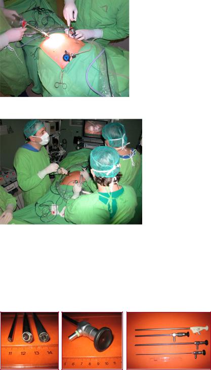
application of sutures at the edges of it. This will prevent the gas to escape (Figure 76. and 77.).
Figure 76. Insertion of the laparoscopic trocar
Figure 77. Clinical setup in a laparoscopic cholecystectomy
7.3.3. Conditions for inspetion of the abdominal cavity, optic
The laparoscope consists of a lens system and an objective. In practice, the Hopkins’ optic is used most frequently. In such an optic the spaces between the lenses-instead of airare filled with the glasses. This increases the light transmission, decreases the light absorption (by almost 70%) and so leads to an improvement in the quaity of the picture (Figure 78.).
Figure 78. Optics
The optical characteristics of the laparoscope are determined by: visual angle, visual field, focal length, and the light loss.
The bigness of the visual angle is marked by the closed angle that is made by the axes of the objective and other lenses of the laparoscope which depends on the direction of optics. The 0°
77
laparoscope provides a straight forward view, and the 30° laparoscope a forward oblique view. The visual field means: ”how wide( or broad) the optic see”.
Light source
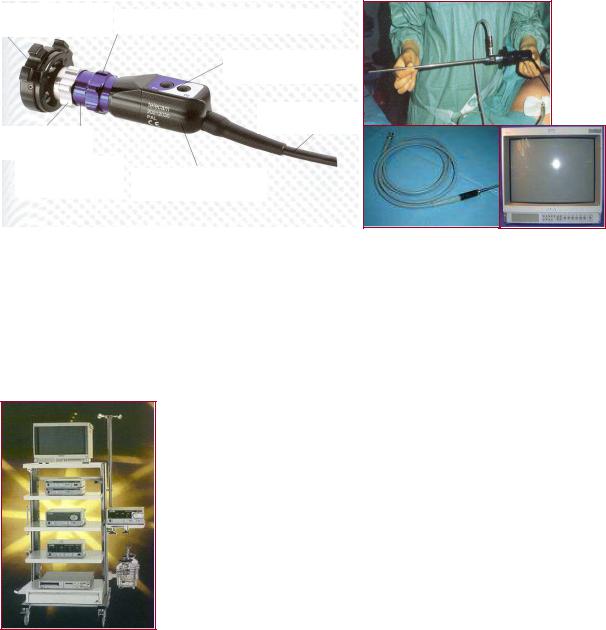
Illumination of the abdominal cavity is of a basic importance for orientation and suitable carrying out of the surgical steps. Currently, a 150–300 W fan-cooled xenon light source is used to provide color-corrected light for extended periods of time without overheating. The illumination is transmitted to the laparoscope via a flexible fiber-optic light guide (180–250 cm long).The camera is connected to the optic.It recieves the arriving picture and transmit it to the monitor (Figure 79. and 80.).
Ocular coupler |
Zoom optics 25-50 mm |
|
Program selector
Connecting
cable
Focusing ring
Ocular coupler
2Video sensor housing
A
B
D
C
Figure 79. Laparoscopic camera (A), Optic and long connected light cable and camera (B), Light cable (C), Monitor (D)
Laparoscopic irrigation/suction device:
It is a necessary device in laparoscopic surgeries. Its central unit is continuously producing a 180 mmHg positiveand a 500 mmHg negative pressure. These effects can be applied into the abdominal cavity with the help of tubes and a valve. As an irrigating solution, we use the warm saline solution.
A
B
F
C
D
E
Figure 80. Laparoscopic tower
A. Monitor, B. Insufflator, C. Light source, D. Electrocautery device, E. Video device, F. Irrigation/suction device
78
Electrocautery devices
They are similar to those used in open conventional surgeries and can be either monoor bipolar. In a monopolar system the circuit of the electric current is made by the active electrode (i.e. hand-held instrument), the patient’s tissue, and the ground pad (i.e. the indifferent electrode). It can possibly cause burn injury of the distant tissues. A bipolar system, in contrast, places the tissue between two electrodes, so the current passes from one electrode to the other through the interposed tissue. In this manner, it is safer than a monopolar system.
Laparoscopic hand instruments
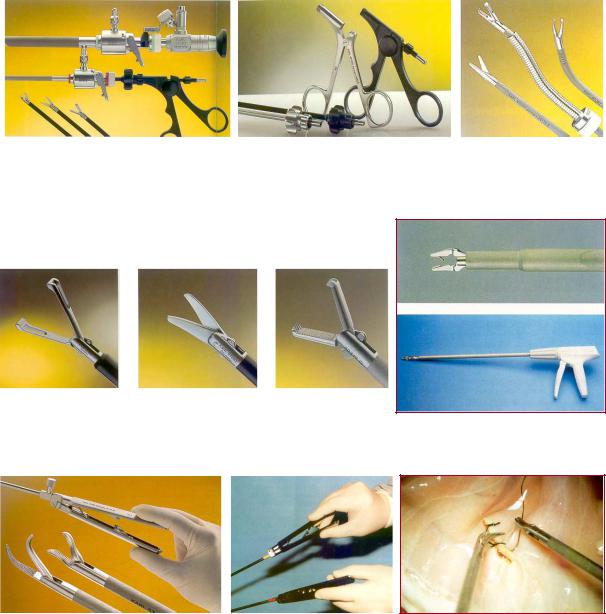
For videoendoscopic surgeries special instruments are needed which are different in sizes, lengths and forms from those used in the open conventional surgeries (Figure 81., 82., and 83.).
A |
|
B |
|
C |
Figure 81. Laparoscopic hand instuments
A. 5-mm and 10-mm ports with instuments, B. Insulated (black one) and non-insulated graspers, C. Flexibel instruments
A |
|
B |
|
C |
|
|
|
|
|
D
Figure 82. Laparoscopic dissector (A), scissors (B), grasper (B), Clip applier with titanium clips (D)
A |
B |
|
C |
|
|
|
|
Figure 83. Laparoscopic needle holders (”parrot” and ”flamingo”) (A), Correct holding of the needle holders (B), Intracorporeal sutures (C)
79
Difficulties of the laparoscopic technique:
•Towdimentional approach and threedimentional activity
•Eye-hand coordination
•Feeling the depth
•Coordinated use of the dominant and non-dominant hands
•Lack of the tactile sensation
•Magnified surgical territory and finer manipulations
•Fulcrum effect
•Limited movement
•New and unusual instruments
•Continuous care of the techniqual equipment
•Increased physical and mental demands
The laparoscopic technique – due to its known benefitsbecame extraordinarily famous. In spite of this, the application of this method is not easy and needs too many practices. Immediately following entry into abdominal cavity you do feel the unusual orientation. The instruments (which are completely different from the usual and accustomed ones) with respect to the characteristic of the optics actually move in a direction opposite to the surgeon’s aims. Even a very simple activity (e.g. knotting) becomes possible only after many hours practices. It is obvious that to become expert in laparoscopic surgery is not possible with performing it on patients. You can get experinces with practising it on a pelvitrainer and -after getting enough experiences - animals.
80