Материал: все о дофамине
Synthesis
Dopamine is synthesized in a restricted set of cell types, mainly neurons and cells in the medulla of the adrenal glands.[18] The primary and minor metabolic pathways respectively are:
Primary: L-Phenylalanine → L-Tyrosine → L-DOPA → Dopamine[15][16]
Minor: L-Phenylalanine → L-Tyrosine → p-Tyramine → Dopamine[15][16][17]
Minor: L-Phenylalanine → m-Tyrosine → m-Tyramine → Dopamine[17][19][20]
The direct precursor of dopamine, L-DOPA, can be synthesized indirectly from the essential amino acid phenylalanine or directly from the non-essential amino acid tyrosine.[21] These amino acids are found in nearly every protein and so are readily available in food, with tyrosine being the most common. Although dopamine is also found in many types of food, it is incapable of crossing the blood–brain barrier that surrounds and protects the brain.[22] It must therefore be synthesized inside the brain to perform its neuronal activity.[22]
L-Phenylalanine is converted into L-tyrosine by the enzyme phenylalanine hydroxylase, with molecular oxygen (O2) and tetrahydrobiopterin as cofactors. L-Tyrosine is converted into L-DOPA by the enzyme tyrosine hydroxylase, with tetrahydrobiopterin, O2, and iron (Fe2+) as cofactors.[21] L-DOPA is converted into dopamine by the enzyme aromatic L-amino acid decarboxylase (also known as DOPA decarboxylase), with pyridoxal phosphate as the cofactor.[21]
Dopamine itself is used as precursor in the synthesis of the neurotransmitters norepinephrine and epinephrine.[21] Dopamine is converted into norepinephrine by the enzyme dopamine β-hydroxylase, with O2 and L-ascorbic acid as cofactors.[21] Norepinephrine is converted into epinephrine by the enzyme phenylethanolamine N-methyltransferase with S-adenosyl-L-methionine as the cofactor.[21]
Some of the cofactors also require their own synthesis.[21] Deficiency in any required amino acid or cofactor can impair the synthesis of dopamine, norepinephrine, and epinephrine.[21]
Degradation
Dopamine is broken down into inactive metabolites by a set of enzymes—monoamine oxidase (MAO), catechol-O-methyl transferase (COMT), and aldehyde dehydrogenase (ALDH), acting in sequence.[23] Both isoforms of monoamine oxidase, MAO-A and MAO-B, effectively metabolize dopamine.[21] Different breakdown pathways exist but the main end-product is homovanillic acid (HVA), which has no known biological activity.[23] From the bloodstream, homovanillic acid is filtered out by the kidneys and then excreted in the urine.[23] The two primary metabolic routes that convert dopamine into HVA are:
Dopamine → DOPAL → DOPAC → HVA – catalyzed by MAO, ALDH, and COMT respectively
Dopamine → 3-Methoxytyramine → HVA – catalyzed by COMT and MAO+ALDH respectively
In clinical research on schizophrenia, measurements of homovanillic acid in plasma have been used to estimate levels of dopamine activity in the brain. A difficulty in this approach however, is separating the high level of plasma homovanillic acid contributed by the metabolism of norepinephrine.[24][25]
Although dopamine is normally broken down by an oxidoreductase enzyme, it is also susceptible to oxidation by direct reaction with oxygen, yielding quinones plus various free radicals as products.[26] The rate of oxidation can be increased by the presence of ferric iron or other factors. Quinones and free radicals produced by autoxidation of dopamine can poison cells, and there is evidence that this mechanism may contribute to the cell loss that occurs in Parkinson's disease and other conditions.[27]
Functions
Cellular effects
Main articles: Dopamine receptor and TAAR1
Primary targets of dopamine in the human brain[1][28] |
||||
Family |
Receptor |
Gene |
Type |
Mechanism |
D1-like |
D1 |
DRD1 |
Gs-coupled. |
Increase intracellular levels of cAMP by activating adenylate cyclase. |
D5 |
DRD5 |
|||
D2-like |
D2 |
DRD2 |
Gi-coupled. |
Decrease intracellular levels of cAMP by inhibiting adenylate cyclase. |
D3 |
DRD3 |
|||
D4 |
DRD4 |
|||
TAAR |
TAAR1 |
TAAR1 |
Gs-coupled. Gq-coupled. |
Increase intracellular levels of cAMP and intracellular calcium concentration. |
Dopamine exerts its effects by binding to and activating cell surface receptors.[18] In humans, dopamine has a high binding affinity at dopamine receptors and human trace amine-associated receptor 1 (hTAAR1).[1][28] In mammals, five subtypes of dopamine receptors have been identified, labeled from D1 to D5.[18] All of them function as metabotropic, G protein-coupled receptors, meaning that they exert their effects via a complex second messenger system.[29] These receptors can be divided into two families, known as D1-like and D2-like.[18] For receptors located on neurons in the nervous system, the ultimate effect of D1-like activation (D1 and D5) can be excitation (via opening of sodium channels) or inhibition (via opening of potassium channels); the ultimate effect of D2-like activation (D2, D3, and D4) is usually inhibition of the target neuron.[29] Consequently, it is incorrect to describe dopamine itself as either excitatory or inhibitory: its effect on a target neuron depends on which types of receptors are present on the membrane of that neuron and on the internal responses of that neuron to the second messenger cAMP.[29] D1 receptors are the most numerous dopamine receptors in the human nervous system; D2 receptors are next; D3, D4, and D5 receptors are present at significantly lower levels.[29]
Storage, release, and reuptake
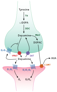
Dopamine processing in a synapse. After release dopamine can either be taken up again by the presynaptic terminal, or broken down by enzymes. TH: tyrosine hydroxylase DOPA: L-DOPA DAT: dopamine transporter DDC: DOPA decarboxylase VMAT: vesicular monoamine transporter 2 MAO: Monoamine oxidase COMT: Catechol-O-methyl transferase HVA: Homovanillic acid
Inside the brain, dopamine functions as a neurotransmitter and neuromodulator, and is controlled by a set of mechanisms common to all monoamine neurotransmitters.[18] After synthesis, dopamine is transported from the cytosol into synaptic vesicles by a solute carrier—a vesicular monoamine transporter, VMAT2.[30] Dopamine is stored in these vesicles until it is ejected into the synaptic cleft. In most cases, the release of dopamine occurs through a process called exocytosis which is caused by action potentials, but it can also be caused by the activity of an intracellular trace amine-associated receptor, TAAR1.[28] TAAR1 is a high-affinity receptor for dopamine, trace amines, and certain substituted amphetamines that is located along membranes in the intracellular milieu of the presynaptic cell;[28] activation of the receptor can regulate dopamine signaling by inducing dopamine reuptake inhibition and efflux as well as by inhibiting neuronal firing through a diverse set of mechanisms.[28][31]
Once in the synapse, dopamine binds to and activates dopamine receptors.[32] These can be postsynaptic dopamine receptors, which are located on dendrites (the postsynaptic neuron), or presynaptic autoreceptors (e.g., the D2sh and presynaptic D3 receptors), which are located on the membrane of an axon terminal (the presynaptic neuron).[18][32] After the postsynaptic neuron elicits an action potential, dopamine molecules quickly become unbound from their receptors. They are then absorbed back into the presynaptic cell, via reuptake mediated either by the dopamine transporter or by the plasma membrane monoamine transporter.[33] Once back in the cytosol, dopamine can either be broken down by a monoamine oxidase or repackaged into vesicles by VMAT2, making it available for future release.[30]
In the brain the level of extracellular dopamine is modulated by two mechanisms: phasic and tonic transmission.[34] Phasic dopamine release, like most neurotransmitter release in the nervous system, is driven directly by action potentials in the dopamine-containing cells.[34] Tonic dopamine transmission occurs when small amounts of dopamine are released without being preceded by presynaptic action potentials.[34] Tonic transmission is regulated by a variety of factors, including the activity of other neurons and neurotransmitter reuptake.[34]
Nervous system
Main articles: Dopaminergic cell groups and Dopaminergic pathways
See also: Hypothalamic–pituitary–prolactin axis
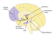
Major dopamine pathways. As part of the reward pathway, dopamine is manufactured in nerve cell bodies located within the ventral tegmental area (VTA) and is released in the nucleus accumbens and the prefrontal cortex. The motor functions of dopamine are linked to a separate pathway, with cell bodies in the substantia nigra that manufacture and release dopamine into the dorsal striatum.
Inside the brain, dopamine plays important roles in executive functions, motor control, motivation, arousal, reinforcement, and reward, as well as lower-level functions including lactation, sexual gratification, and nausea. The dopaminergic cell groups and pathways make up the dopamine system which is neuromodulatory.
Dopaminergic neurons (dopamine-producing nerve cells) are comparatively few in number—a total of around 400,000 in the human brain[35]—and their cell bodies are confined in groups to a few relatively small brain areas.[36] However their axons project to many other brain areas, and they exert powerful effects on their targets.[36] These dopaminergic cell groups were first mapped in 1964 by Annica Dahlström and Kjell Fuxe, who assigned them labels starting with the letter "A" (for "aminergic").[37] In their scheme, areas A1 through A7 contain the neurotransmitter norepinephrine, whereas A8 through A14 contain dopamine. The dopaminergic areas they identified are the substantia nigra (groups 8 and 9); the ventral tegmental area (group 10); the posterior hypothalamus (group 11); the arcuate nucleus (group 12); the zona incerta (group 13) and the periventricular nucleus (group 14).[37]
The substantia nigra is a small midbrain area that forms a component of the basal ganglia. This has two parts—an input area called the pars compacta and an output area the pars reticulata. The dopaminergic neurons are found mainly in the pars compacta (cell group A8) and nearby (group A9).[36] In humans, the projection of dopaminergic neurons from the substantia nigra pars compacta to the dorsal striatum, termed the nigrostriatal pathway, plays a significant role in the control of motor function and in learning new motor skills.[38] These neurons are especially vulnerable to damage, and when a large number of them die, the result is a parkinsonian syndrome.[39]
The ventral tegmental area (VTA) is another midbrain area. The most prominent group of VTA dopaminergic neurons projects to the prefrontal cortex via the mesocortical pathway and another smaller group projects to the nucleus accumbens via the mesolimbic pathway. Together, these two pathways are collectively termed the mesocorticolimbic projection.[36][38] The VTA also sends dopaminergic projections to the amygdala, cingulate gyrus, hippocampus, and olfactory bulb.[36][38] Mesocorticolimbic neurons play a central role in reward and other aspects of motivation.[38] Accumulating literature shows that dopamine also plays a crucial role in aversive learning through its effects on a number of brain regions.[40][41][42]
The posterior hypothalamus has dopamine neurons that project to the spinal cord, but their function is not well established.[43] There is some evidence that pathology in this area plays a role in restless legs syndrome, a condition in which people have difficulty sleeping due to an overwhelming compulsion to constantly move parts of the body, especially the legs.[43]
The arcuate nucleus and the periventricular nucleus of the hypothalamus have dopamine neurons that form an important projection—the tuberoinfundibular pathway which goes to the pituitary gland, where it influences the secretion of the hormone prolactin.[44] Dopamine is the primary neuroendocrine inhibitor of the secretion of prolactin from the anterior pituitary gland.[44] Dopamine produced by neurons in the arcuate nucleus is secreted into the hypophyseal portal system of the median eminence, which supplies the pituitary gland.[44] The prolactin cells that produce prolactin, in the absence of dopamine, secrete prolactin continuously; dopamine inhibits this secretion.[44] In the context of regulating prolactin secretion, dopamine is occasionally called prolactin-inhibiting factor, prolactin-inhibiting hormone, or prolactostatin.[44]
The zona incerta, grouped between the arcuate and periventricular nuclei, projects to several areas of the hypothalamus, and participates in the control of gonadotropin-releasing hormone, which is necessary to activate the development of the male and female reproductive systems, following puberty.[44]
An additional group of dopamine-secreting neurons is found in the retina of the eye.[45] These neurons are amacrine cells, meaning that they have no axons.[45] They release dopamine into the extracellular medium, and are specifically active during daylight hours, becoming silent at night.[45] This retinal dopamine acts to enhance the activity of cone cells in the retina while suppressing rod cells—the result is to increase sensitivity to color and contrast during bright light conditions, at the cost of reduced sensitivity when the light is dim.[45]
Basal ganglia
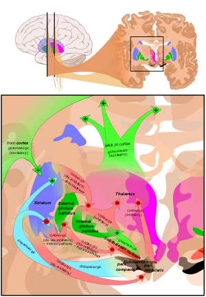
Main circuits of the basal ganglia. The dopaminergic pathway from the substantia nigra pars compacta to the striatum is shown in light blue.
The largest and most important sources of dopamine in the vertebrate brain are the substantia nigra and ventral tegmental area.[36] These structures are closely related to each other and functionally similar in many respects.[36] Both are components of the mid brain.[36] The largest component of the basal ganglia is the striatum.[46] The substantia nigra sends a dopaminergic projection to the dorsal striatum, while the ventral tegmental area sends a similar type of dopaminergic projection to the ventral striatum.[36]
Progress in understanding the functions of the basal ganglia has been slow.[46] The most popular hypotheses, broadly stated, propose that the basal ganglia play a central role in action selection.[47] The action selection theory in its simplest form proposes that when a person or animal is in a situation where several behaviors are possible, activity in the basal ganglia determines which of them is executed, by releasing that response from inhibition while continuing to inhibit other motor systems that if activated would generate competing behaviors.[48] Thus the basal ganglia, in this concept, are responsible for initiating behaviors, but not for determining the details of how they are carried out. In other words, they essentially form a decision-making system.[48]
The basal ganglia can be divided into several sectors, and each is involved in controlling particular types of actions.[49] The ventral sector of the basal ganglia (containing the ventral striatum and ventral tegmental area) operates at the highest level of the hierarchy, selecting actions at the whole-organism level.[48] The dorsal sectors (containing the dorsal striatum and substantia nigra) operate at lower levels, selecting the specific muscles and movements that are used to implement a given behavior pattern.[49]
Dopamine contributes to the action selection process in at least two important ways. First, it sets the "threshold" for initiating actions.[47] The higher the level of dopamine activity, the lower the impetus required to evoke a given behavior.[47] As a consequence, high levels of dopamine lead to high levels of motor activity and impulsive behavior; low levels of dopamine lead to torpor and slowed reactions.[47] Parkinson's disease, in which dopamine levels in the substantia nigra circuit are greatly reduced, is characterized by stiffness and difficulty initiating movement—however, when people with the disease are confronted with strong stimuli such as a serious threat, their reactions can be as vigorous as those of a healthy person.[50] In the opposite direction, drugs that increase dopamine release, such as cocaine or amphetamine, can produce heightened levels of activity, including, at the extreme, psychomotor agitation and stereotyped movements.[51]
The second important effect of dopamine is as a "teaching" signal.[47] When an action is followed by an increase in dopamine activity, the basal ganglia circuit is altered in a way that makes the same response easier to evoke when similar situations arise in the future.[47] This is a form of operant conditioning, in which dopamine plays the role of a reward signal.[48]
Reward
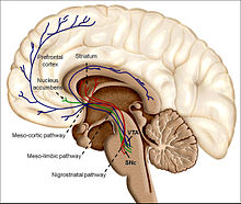
Illustration of dopaminergic reward structures
In the language used to discuss the reward system, reward is the attractive and motivational property of a stimulus that induces appetitive behavior (also known as approach behavior) and consummatory behavior.[52] A rewarding stimulus is one that can induce the organism to approach it and choose to consume it.[52] Pleasure, learning (e.g., classical and operant conditioning), and approach behavior are the three main functions of reward.[52] As an aspect of reward, pleasure provides a definition of reward;[52] however, while all pleasurable stimuli are rewarding, not all rewarding stimuli are pleasurable (e.g., extrinsic rewards like money).[52][53] The motivational or desirable aspect of rewarding stimuli is reflected by the approach behavior that they induce, whereas the pleasure from intrinsic rewards results from consuming them after acquiring them.[52] A neuropsychological model which distinguishes these two components of an intrinsically rewarding stimulus is the incentive salience model, where "wanting" or desire (less commonly, "seeking"[54]) corresponds to appetitive or approach behavior while "liking" or pleasure corresponds to consummatory behavior.[52][4][55] In human drug addicts, "wanting" becomes dissociated with "liking" as the desire to use an addictive drug increases, while the pleasure obtained from consuming it decreases due to drug tolerance.[4]