Материал: все о дофамине
Psychosis and antipsychotic drugs
Main article: Psychosis
Psychiatrists in the early 1950s discovered that a class of drugs known as typical antipsychotics (also known as major tranquilizers), were often effective at reducing the psychotic symptoms of schizophrenia.[112] The introduction of the first widely used antipsychotic, chlorpromazine (Thorazine), in the 1950s, led to the release of many patients with schizophrenia from institutions in the years that followed.[112] By the 1970s researchers understood that these typical antipsychotics worked as antagonists on the D2 receptors.[112][113] This realization led to the so-called dopamine hypothesis of schizophrenia, which postulates that schizophrenia is largely caused by hyperactivity of brain dopamine systems.[114] The dopamine hypothesis drew additional support from the observation that psychotic symptoms were often intensified by dopamine-enhancing stimulants such as methamphetamine, and that these drugs could also produce psychosis in healthy people if taken in large enough doses.[114] In the following decades other atypical antipsychotics that had fewer serious side effects were developed.[112] Many of these newer drugs do not act directly on dopamine receptors, but instead produce alterations in dopamine activity indirectly.[115] These drugs were also used to treat other psychoses.[112] Antipsychotic drugs have a broadly suppressive effect on most types of active behavior, and particularly reduce the delusional and agitated behavior characteristic of overt psychosis.[113]
Later observations, however, have caused the dopamine hypothesis to lose popularity, at least in its simple original form.[114] For one thing, patients with schizophrenia do not typically show measurably increased levels of brain dopamine activity.[114] Even so, many psychiatrists and neuroscientists continue to believe that schizophrenia involves some sort of dopamine system dysfunction.[112] As the "dopamine hypothesis" has evolved over time, however, the sorts of dysfunctions it postulates have tended to become increasingly subtle and complex.[112]
Psychopharmacologist Stephen M. Stahl suggested in a review of 2018 that in many cases of psychosis, including schizophrenia, three interconnected networks based on dopamine, serotonin, and glutamate – each on its own or in various combinations – contributed to an overexcitation of dopamine D2 receptors in the ventral striatum.[116]
Attention deficit hyperactivity disorder
Altered dopamine neurotransmission is implicated in attention deficit hyperactivity disorder (ADHD), a condition associated with impaired cognitive control, in turn leading to problems with regulating attention (attentional control), inhibiting behaviors (inhibitory control), and forgetting things or missing details (working memory), among other problems.[117] There are genetic links between dopamine receptors, the dopamine transporter, and ADHD, in addition to links to other neurotransmitter receptors and transporters.[118] The most important relationship between dopamine and ADHD involves the drugs that are used to treat ADHD.[119] Some of the most effective therapeutic agents for ADHD are psychostimulants such as methylphenidate (Ritalin, Concerta) and amphetamine (Evekeo, Adderall, Dexedrine), drugs that increase both dopamine and norepinephrine levels in the brain.[119] The clinical effects of these psychostimulants in treating ADHD are mediated through the indirect activation of dopamine and norepinephrine receptors, specifically dopamine receptor D1 and adrenoceptor α2, in the prefrontal cortex.[117][120][121]
Pain
Dopamine plays a role in pain processing in multiple levels of the central nervous system including the spinal cord, periaqueductal gray, thalamus, basal ganglia, and cingulate cortex.[122] Decreased levels of dopamine have been associated with painful symptoms that frequently occur in Parkinson's disease.[122] Abnormalities in dopaminergic neurotransmission also occur in several painful clinical conditions, including burning mouth syndrome, fibromyalgia, and restless legs syndrome.[122]
Nausea
Nausea and vomiting are largely determined by activity in the area postrema in the medulla of the brainstem, in a region known as the chemoreceptor trigger zone.[123] This area contains a large population of type D2 dopamine receptors.[123] Consequently, drugs that activate D2 receptors have a high potential to cause nausea.[123] This group includes some medications that are administered for Parkinson's disease, as well as other dopamine agonists such as apomorphine.[124] In some cases, D2-receptor antagonists such as metoclopramide are useful as anti-nausea drugs.[123]
Comparative biology and evolution
Microorganisms
There are no reports of dopamine in archaea, but it has been detected in some types of bacteria and in the protozoan called Tetrahymena.[125] Perhaps more importantly, there are types of bacteria that contain homologs of all the enzymes that animals use to synthesize dopamine.[126] It has been proposed that animals derived their dopamine-synthesizing machinery from bacteria, via horizontal gene transfer that may have occurred relatively late in evolutionary time, perhaps as a result of the symbiotic incorporation of bacteria into eukaryotic cells that gave rise to mitochondria.[126]
Animals
Dopamine is used as a neurotransmitter in most multicellular animals.[127] In sponges there is only a single report of the presence of dopamine, with no indication of its function;[128] however, dopamine has been reported in the nervous systems of many other radially symmetric species, including the cnidarian jellyfish, hydra and some corals.[129] This dates the emergence of dopamine as a neurotransmitter back to the earliest appearance of the nervous system, over 500 million years ago in the Cambrian Period. Dopamine functions as a neurotransmitter in vertebrates, echinoderms, arthropods, molluscs, and several types of worm.[130][131]
In every type of animal that has been examined, dopamine has been seen to modify motor behavior.[127] In the model organism, nematode Caenorhabditis elegans, it reduces locomotion and increases food-exploratory movements; in flatworms it produces "screw-like" movements; in leeches it inhibits swimming and promotes crawling. Across a wide range of vertebrates, dopamine has an "activating" effect on behavior-switching and response selection, comparable to its effect in mammals.[127][132]
Dopamine has also consistently been shown to play a role in reward learning, in all animal groups.[127] As in all vertebrates – invertebrates such as roundworms, flatworms, molluscs and common fruit flies can all be trained to repeat an action if it is consistently followed by an increase in dopamine levels.[127] In fruit flies, distinct elements for reward learning suggest a modular structure to the insect reward processing system that broadly parallels that the mammalian one.[133] For example, dopamine regulates short- and long-term learning in monkeys;[134] in fruit flies, different groups of dopamine neurons mediate reward signals for short- and long-term memories.[135]
It had long been believed that arthropods were an exception to this with dopamine being seen as having an adverse effect. Reward was seen to be mediated instead by octopamine, a neurotransmitter closely related to norepinephrine.[136] More recent studies, however, have shown that dopamine does play a part in reward learning in fruit flies. It has also been found that the rewarding effect of octopamine is due to its activating a set of dopaminergic neurons not previously accessed in the research.[136]
Plants
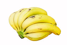
Dopamine can be found in the peel and fruit pulp of bananas.
Many plants, including a variety of food plants, synthesize dopamine to varying degrees.[137] The highest concentrations have been observed in bananas—the fruit pulp of red and yellow bananas contains dopamine at levels of 40 to 50 parts per million by weight.[137] Potatoes, avocados, broccoli, and Brussels sprouts may also contain dopamine at levels of 1 part per million or more; oranges, tomatoes, spinach, beans, and other plants contain measurable concentrations less than 1 part per million.[137] The dopamine in plants is synthesized from the amino acid tyrosine, by biochemical mechanisms similar to those that animals use.[137] It can be metabolized in a variety of ways, producing melanin and a variety of alkaloids as byproducts.[137] The functions of plant catecholamines have not been clearly established, but there is evidence that they play a role in the response to stressors such as bacterial infection, act as growth-promoting factors in some situations, and modify the way that sugars are metabolized. The receptors that mediate these actions have not yet been identified, nor have the intracellular mechanisms that they activate.[137]
Dopamine consumed in food cannot act on the brain, because it cannot cross the blood–brain barrier.[22] However, there are also a variety of plants that contain L-DOPA, the metabolic precursor of dopamine.[138] The highest concentrations are found in the leaves and bean pods of plants of the genus Mucuna, especially in Mucuna pruriens (velvet beans), which have been used as a source for L-DOPA as a drug.[139] Another plant containing substantial amounts of L-DOPA is Vicia faba, the plant that produces fava beans (also known as "broad beans"). The level of L-DOPA in the beans, however, is much lower than in the pod shells and other parts of the plant.[140] The seeds of Cassia and Bauhinia trees also contain substantial amounts of L-DOPA.[138]
In a species of marine green algae Ulvaria obscura, a major component of some algal blooms, dopamine is present in very high concentrations, estimated at 4.4% of dry weight. There is evidence that this dopamine functions as an anti-herbivore defense, reducing consumption by snails and isopods.[141]
As a precursor for melanin
Melanins are a family of dark-pigmented substances found in a wide range of organisms.[142] Chemically they are closely related to dopamine, and there is a type of melanin, known as dopamine-melanin, that can be synthesized by oxidation of dopamine via the enzyme tyrosinase.[142] The melanin that darkens human skin is not of this type: it is synthesized by a pathway that uses L-DOPA as a precursor but not dopamine.[142] However, there is substantial evidence that the neuromelanin that gives a dark color to the brain's substantia nigra is at least in part dopamine-melanin.[143]
Dopamine-derived melanin probably appears in at least some other biological systems as well. Some of the dopamine in plants is likely to be used as a precursor for dopamine-melanin.[144] The complex patterns that appear on butterfly wings, as well as black-and-white stripes on the bodies of insect larvae, are also thought to be caused by spatially structured accumulations of dopamine-melanin.[145]
History and development
Main article: History of catecholamine research
Dopamine was first synthesized in 1910 by George Barger and James Ewens at Wellcome Laboratories in London, England[146] and first identified in the human brain by Katharine Montagu in 1957. It was named dopamine because it is a monoamine whose precursor in the Barger-Ewens synthesis is 3,4-dihydroxyphenylalanine (levodopa or L-DOPA). Dopamine's function as a neurotransmitter was first recognized in 1958 by Arvid Carlsson and Nils-Åke Hillarp at the Laboratory for Chemical Pharmacology of the National Heart Institute of Sweden.[147] Carlsson was awarded the 2000 Nobel Prize in Physiology or Medicine for showing that dopamine is not only a precursor of norepinephrine (noradrenaline) and epinephrine (adrenaline), but is also itself a neurotransmitter.[148]
Polydopamine
Research motivated by adhesive polyphenolic proteins in mussels led to the discovery in 2007 that a wide variety of materials, if placed in a solution of dopamine at slightly basic pH, will become coated with a layer of polymerized dopamine, often referred to as polydopamine.[149][150] This polymerized dopamine forms by a spontaneous oxidation reaction, and is formally a type of melanin.[151] Synthesis usually involves reaction of dopamine hydrochloride with Tris as a base in water. The structure of polydopamine is unknown.[150]
Polydopamine coatings can form on objects ranging in size from nanoparticles to large surfaces.[151] Polydopamine layers have chemical properties that have the potential to be extremely useful, and numerous studies have examined their possible applications.[151] At the simplest level, they can be used for protection against damage by light, or to form capsules for drug delivery.[151] At a more sophisticated level, their adhesive properties may make them useful as substrates for biosensors or other biologically active macromolecules.[151]
TAAR1
From Wikipedia, the free encyclopedia
(Redirected from Human trace amine-associated receptor 1)
Jump to navigationJump to search
TAAR1 |
|||||||
|
|||||||
Identifiers |
|||||||
Aliases |
TAAR1, TA1, TAR1, TRAR1, trace amine associated receptor 1, Trace amine receptor |
||||||
External IDs |
OMIM: 609333 MGI: 2148258 HomoloGene: 24938 GeneCards: TAAR1 |
||||||
|
|||||||
|
|||||||
|
|||||||
Orthologs |
|||||||
Species |
Human |
Mouse |
|
||||
Entrez |
|
|
|
||||
Ensembl |
|
|
|
||||
UniProt |
|
|
|
||||
RefSeq (mRNA) |
|
|
|
||||
RefSeq (protein) |
|
|
|
||||
Location (UCSC) |
Chr 6: 132.64 – 132.66 Mb |
n/a |
|
||||
PubMed search |
[2] |
[3] |
|
||||
Wikidata |
|||||||
|
|||||||
Trace amine-associated receptor 1 (TAAR1) is a trace amine-associated receptor (TAAR) protein that in humans is encoded by the TAAR1 gene.[4] TAAR1 is an intracellular amine-activated Gs-coupled and Gq-coupled G protein-coupled receptor (GPCR) that is primarily expressed in several peripheral organs and cells (e.g., the stomach, small intestine, duodenum, and white blood cells), astrocytes, and in the intracellular milieu within the presynaptic plasma membrane (i.e., axon terminal) of monoamine neurons in the central nervous system (CNS).[5][6][7][8] TAAR1 was discovered in 2001 by two independent groups of investigators, Borowski et al. and Bunzow et al.[9][10] TAAR1 is one of six functional human trace amine-associated receptors, which are so named for their ability to bind endogenous amines that occur in tissues at trace concentrations.[11][12] TAAR1 plays a significant role in regulating neurotransmission in dopamine, norepinephrine, and serotonin neurons in the CNS;[6][11] it also affects immune system and neuroimmune system function through different mechanisms.[13][14][15][16]
TAAR1 is a high-affinity receptor for amphetamine, methamphetamine, dopamine, and trace amines which mediates some of their cellular effects in monoamine neurons within the central nervous system.[6][11]
The primary endogenous ligands of the human TAAR1 (hTAAR1) receptor, by rank order of potency, are: tyramine > β-phenethylamine > dopamine = octopamine.[5]
Contents
1Discovery
2Structure
3Gene
4Tissue distribution
4.1Location within neurons
4.2Receptor oligomers
5Ligands
5.1Agonists
5.1.1Trace amines
5.1.2Thyronamines
5.1.3Synthetic
5.2Partial agonists
5.3Inverse agonists
5.4Neutral antagonists
6Function
6.1Monoaminergic systems
6.2Immune system
7Clinical significance
7.1Research
8Notes
9References
10External links
Discovery[edit]
TAAR1 was discovered independently by Borowski et al. and Bunzow et al. in 2001. To find the genetic variants responsible for TAAR1 synthesis, they used mixtures of oligonucleotides with sequences related to G protein-coupled receptors (GPCRs) of serotonin and dopamine to discover novel DNA sequences in rat genomic DNA and cDNA, which they then amplified and cloned. The resulting sequence was not found in any database and coded for TAAR1.[9][10] Later, the functional role of TAAR1 and other receptors from this family was studied by Raul Gainetdinov and his colleagues.[17]
Structure[edit]
TAAR1 shares structural similarities with the class A rhodopsin GPCR subfamily.[10] It has 7 transmembrane domains with short N and C terminal extensions.[18] TAAR1 is 62–96% identical with TAARs2-15, which suggests that the TAAR subfamily has recently evolved; while at the same time, the low degree of similarity between TAAR1 orthologues suggests that they are rapidly evolving.[9] TAAR1 shares a predictive peptide motif with all other TAARs. This motif overlaps with transmembrane domain VII, and its identity is NSXXNPXX[Y,H]XXX[Y,F]XWF. TAAR1 and its homologues have ligand pocket vectors that utilize sets of 35 amino acids known to be involved directly in receptor-ligand interaction.[12]