Материал: 100_Cases_in_Clinical_Medicine

ANSWER 23
This patient has diarrhoea and weight loss due to giardiasis. It is important to take a careful history in a patient with diarrhoea. An acute infective diarrhoea develops soon after ingestion of the offending food. The commonest infective causes of infective gastroenteritis in the UK are viruses such as rotaviruses, or the bacteria Campylobacter jejuni or Salmonella enteritidis. Staphylococcal food poisoning occurs within a few hours and typically presents abruptly and may be severe but short-lived. Campylobacter, Salmonella and Shigella cause more severe symptoms than viral gastroenteritis. The incubation period for giardiasis is typically about 2 weeks, but varies from 3 days to 6 weeks. Giardia lamblia infects the small intestine and causes a watery, yellow, foul-smelling diarrhoea. Loss of appetite, nausea and weight loss are common. Symptoms usually improve after 2–3 weeks, but can persist, in some cases causing lactose intolerance.
The history should try to distinguish between the smalland large-bowel origin of the diarrhoea. Large-bowel diarrhoea tends to be maximal in the morning, pain is relieved by defaecation, and blood and mucus may be present. By contrast diarrhoea of small-bowel origin does not occur at any particular time, and pain is not helped by defaecation. Typically a pale fatty stool without blood or mucus occurs in small-bowel disease. Other pathogens which cause small-bowel diarrhoea include Campylobacter, rotavirus, Cryptosporidia and Strongyloides. If small-bowel-type diarrhoea persists, other non-infective causes of malabsorption should be considered such as tropical sprue, coeliac disease, and chronic pancreatitis.
Giardia lamblia occurs worldwide especially in the tropics but also is endemic in Russia, and infection occurs commonly in visitors to St Petersburg. Poor sanitation and untreated water supplies are important factors in transmission. Outbreaks can occur in residents of nursing homes, and giardiasis is a common cause of diarrhoea in homosexuals.
Diagnosis is made by finding cysts of the parasite in stool samples. If stool samples are negative, cysts can be found on jejunal biopsy or by sampling duodenal fluid by asking the patient to swallow the Enterotest capsule. Treatment is with a course of metronidazole. Ideally a stool sample should be examined 6 weeks after treatment to ensure the parasite has been eradicated. Alcohol, dairy products and spices should be avoided during the recovery period.
If no infective cause had been found for this man’s diarrhoea and weight loss, further investigations would have been necessary to exclude causes such as malignancy or thyrotoxicosis.
KEY POINTS
•Stool samples should be sent in a patient returning from abroad with diarrhoea.
•Always take an accurate history of recent foreign travel in a patient with diarrhoea.
•Attempt to distinguish clinically between smalland large-bowel origin of the diarrhoea.
•Giardiasis is a common cause of traveller’s diarrhoea.
64
CASE 24: SHORTNESS OF BREATH ON EXERTION
A 23-year-old student presents to her general practitioner (GP) complaining of shortness of breath on exertion. This has developed over the past 10 days, and she is now breathless after walking 50 yards. About 2 weeks ago she had a flu-like illness with generalized muscle aches and fever. She feels extremely tired and has noticed palpitations in association with her breathlessness. In addition she has some discomfort in her anterior chest which is worse on inspiration. Previously she has been extremely fit with no significant past medical history. There is no recent history of foreign travel. She denies substance abuse.
Examination
On examination, her temperature is 37.5°C. Her pulse rate is 120/min and regular. Blood pressure is 90/70 mmHg. Jugular venous pressure is raised at 8 cm. On auscultation there is a gallop rhythm, with a third heart sound. Examination of her chest is unremarkable. Pressure over the sternum causes discomfort. Abdominal and neurological examination is normal.
INVESTIGATIONS
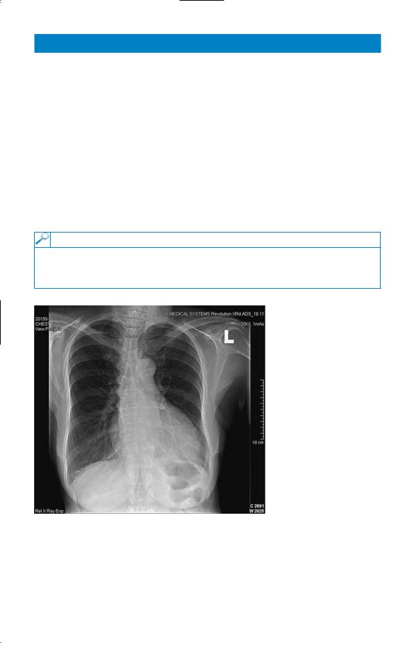
The GP sends the student to the emergency department where an electrocardiogram (ECG) and chest X-ray are performed. The ECG shows T-wave flattening globally. The chest X-ray is shown in Fig. 24.1.
Figure 24.1 Chest X-ray.
Questions
•What is the likely diagnosis?
•How would you further investigate and manage this patient?
65

ANSWER 24
This patient has viral myocarditis due to Coxsackie B virus. Viruses which can cause myocarditis include Coxsackie B and A, echovirus, adenovirus, influenza, varicella, polio, mumps, rabies, viral hepatitis, rubella, Epstein–Barr virus (EBV), cytomegalovirus (CMV), and herpes simplex virus. Myocarditis may occur during a bacteraemia or fungaemia. Rickettsia and diptheria can cause myocarditis. In rural South America acute infection with the protozoan Trypanosoma cruzi causes fever, myocarditis and hepatosplenomegaly, and 10–30 years later this can lead to cardiac failure and conduction system defects (Chagas’ disease). Cocaine abuse can cause myocarditis, and sudden death. Profound hypocalcaemia, hypophosphataemia, and hypomagnaesaemia can all cause myocardial depression.
The clinical picture of myocarditis is non-specific, but common symptoms include myalgia, fatigue, shortness of breath, pericardial pain and palpitations. The prodromal flu-like illness is suggestive of this condition. The main clinical signs are those of cardiac failure. Patients usually have a marked sinus tachycardia disproportionate to the slight fever. ECG usually shows ST segment and T-wave abnormalities. There may be atrial or, more commonly, ventricular arrhythmias or signs of conducting system defects. Chest X-ray may be normal if the myocarditis is mild, but if there is cardiac failure there will be cardiomegaly and pulmonary congestion. The differential diagnoses in this case include hypertrophic cardiomyopathy, pericarditis and myocardial ischaemia.
Echocardiography should be performed to confirm the diagnosis. Echocardiographic changes may be focal affecting only the right or left ventricle, or global. There is poor contractility of the myocardium. Cardiac enzymes such as creatine kinase are raised. An endomyocardial biopsy is performed as soon as possible, and will show evidence of myocardial necrosis. Paired serum samples should be taken for antibody titres to Coxsackie B and mumps. Coxsackie virus can be cultured from the throat, stool, blood, myocardium or pericardial fluid.
Bed rest is the treatment for the period of acute viral myocarditis. Diuretics and angiotensinconverting enzyme (ACE) inhibitors are used to treat cardiac failure. There is controversy over treatment with corticosteroids. Corticosteroids tend to be used in patients with a short history, a positive endomyocardial biopsy, and the most severe disease. Most cases are benign and self-limiting, and cardiac function will return to normal. However a minority will develop permanent cardiac damage leading to a dilated cardiomyopathy. Definitive treatment may then involve cardiac transplantation.
KEY POINTS
•The features in favour of the diagnosis of viral myocarditis include the young age of the patient, the preceding acute febrile illness and subsequently the raised serum antibody titres to Coxsackie B.
•It is important to take a history of foreign travel and substance abuse
•Outcome in adults is generally good, but a proportion of patients will develop a dilated cardiomyopathy.
66
This page intentionally left blank

CASE 25: FEVER AND SHORTNESS OF BREATH
History
A 62-year-old man presents to the emergency department complaining of shortness of breath. Four days prior to presentation he felt unwell and complained of muscle aches and headache. He started having rigors and his wife measured his temperature as 39°C. They thought that he had influenza. However his symptoms worsened, and by the day of presentation he was complaining of a dry cough and marked shortness of breath. He had also become confused, and started having diarrhoea. There is no significant past medical history. He is a non-smoker and drinks 20 units of alcohol a week. Ten days prior to admission he had returned from a holiday in Spain.
Examination
On examination the patient looks unwell, dehydrated and flushed. His temperature is 39.5°C. He has central cyanosis. His pulse rate is 120/min and blood pressure 146/72 mmHg. His respiratory rate is 32/min. His trachea is central and chest expansion is symmetrical. Percussion is reduced, and auscultation reveals bilateral crackles and bronchial breathing in both lower zones posteriorly. His abdomen is diffusely tender but there is no rigidity or guarding. He is disorientated in time, place and person.
Blood tests, arterial blood gases on air, urinalysis and chest X-ray (Fig. 25.1) are shown below.
INVESTIGATIONS
|
|
Normal |
Haemoglobin |
15.3 g/dL |
13.3–17.7 g/dL |
White cell count |
10.3 % 109/L |
3.9–10.6 % 109/L |
Neutrophils |
8.9 % 109/L |
1.8–7.7 % 109/L |
Lymphocytes |
0.4 % 109/L |
0.6–4.8 % 109/L |
Platelets |
143 % 109/L |
150–440 % 109/L |
Sodium |
124 mmol/L |
135–145 mmol/L |
Potassium |
4.4 mmol/L |
3.5–5.0 mmol/L |
Urea |
14.4 mmol/L |
2.5–6.7 mmol |
Creatinine |
178 &mol/L |
70–120 &mol/L |
Glucose |
7.7 mmol/L |
4.0–6.0 mmol/L |
Calcium |
1.88 mmol/L |
2.12–2.65 mmol/L |
Phosphate |
1.2 mmol/L |
0.8–1.45 mmol/L |
C-reactive protein (CRP) |
256 mg/L |
!5 mg/L |
Arterial blood gases on air |
|
|
pH |
7.38 |
7.38–7.44 |
pCO2 |
2.7 kPa |
4.7–6.0 kPa |
pO2 |
6.3 kPa |
12.0–14.5 kPa |
Urinalysis: '' blood; '' protein |
|
|
68