Материал: 100_Cases_in_Clinical_Medicine
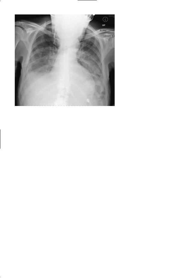
Figure 25.1 Chest X-ray.
Questions
•What is the likely diagnosis?
•How would you further investigate and manage this patient?
69

ANSWER 25
This man has Legionella pneumophila pneumonia. Community-acquired pneumonia is most commonly caused by Streptococcus pneumoniae or Haemophilus influenzae, but atypical pneumonias account for about 5–15 per cent of cases. The 4-day prodromal illness is typical of Legionella pneumonia (2–10 days) compared to pneumococcal pneumonia which tends to present abruptly with fever and shortness of breath. Legionella infection presents with malaise, myalgia, headache and fever. Patients may develop diarrhoea and abdominal pain. As the illness progresses the patient develops a dry cough, chest pain, shortness of breath and acute confusion. Other potential complications include nephritis, endocarditis and myocarditis. On examination, the patient is usually dehydrated, tachycardic and tachypnoeic with widespread rhonchi and crackles. The diffuse infiltrates on chest X-ray suggest atypical pneumonia, whereas a lobar pattern tends to occur with streptococcal pneumonia. Hyponatraemia occurs in cases of severe pneumonia and is a poor prognostic factor. Hypocalcaemia is another distinctive biochemical abnormality in this condition. Confusion and raised urea are markers of severity. The high CRP is consistent with a severe infection, and the lymphopenia is a clue to the fact that this patient has an atypical pneumonia. The patient’s arterial blood gases showed marked hypoxia. This patient presumably acquired his infection while on holiday in Spain. Legionella outbreaks have often been due to infected water tanks in warm climates in institutions such as hotels and hospitals.
This man is acutely unwell and needs to be admitted to a high-dependency unit. He needs to receive high concentration of inspired oxygen, and also intravenous fluids to correct his dehydration. He may require mechanical ventilation. He should be started on intravenous antibiotics. These should cover the common community-acquired pneumonias until the precise microbiological diagnosis is obtained and the antibiotics can then be rationalized. A standard combination is cefuroxime and clarithromycin. Blood cultures should be sent, and blood sent to screen for antibodies to atypical organisms such as Legionella, Mycoplasma, Chlamydia psittaci and influenza. Ten to fourteen days later a further blood sample should be sent and a fourfold rise in antibody titre is evidence of current infection. A faster diagnosis is made by testing broncheoalveolar lavage fluid, blood and urine for the presence of
Legionella antigen.
KEY POINTS
•Legionella is one of the atypical causes of pneumonia.
•It should be suspected if there is an outbreak in an institution, or if a case of pneumonia fails to respond to antibiotics.
•Legionella pneumonia has a 2–10-day prodromal illness.
•Lymphopenia, evidence of nephritis, and a diffuse pattern of infiltrates on chest X-ray are other clues to the diagnosis.
70

CASE 26: LOIN PAIN AND HAEMATURIA
History
A 46-year-old woman presents to the emergency department with a 2-day history of right-sided loin pain and macroscopic haematuria. The pain is continuous and dull in character. Over the past 10 years she has had previous episodes of loin pain which have occurred on both sides and resolved spontaneously over a few days. She has never passed any stones. She was noted to be mildly hypertensive during her three pregnancies. She has no other significant medical history. Her father died of a subarachnoid haemorrhage, aged 48 years. Her father’s brother has had a kidney transplant. She has no siblings. Her three children, aged 17, 14 and 10 years, are well. She works as a teacher and neither smokes nor drinks alcohol.
Examination
On examination she is afebrile. Her pulse is regular at 76/min and her blood pressure is 135/105 mmHg. Examination of the cardiovascular and respiratory systems is otherwise unremarkable. On palpation of her abdomen, ballottable masses are palpable in each flank. The right-sided mass is tender to palpation. Percussion note is resonant over the masses. Neurological examination is normal. Funduscopy shows arteriovenous nipping and silverwiring of the retinal vessels.
INVESTIGATIONS
|
|
Normal |
Haemoglobin |
14.3 g/dL |
11.7–15.7 g/dL |
White cell count |
5.2 % 109/L |
3.5–11.0 % 109/L |
Platelets |
206 % 109/L |
150–440 % 109/L |
Sodium |
138 mmol/L |
135–145 mmol/L |
Potassium |
4.3 mmol/L |
3.5–5.0 mmol/L |
Urea |
10.2 mmol/L |
2.5–6.7 mmol/L |
Creatinine |
146 &mol/L |
70–120 &mol/L |
Albumin |
42 g/L |
35–50 g/L |
Urinalysis: ' protein; ''' blood |
|
|
Urine microscopy: #200 red cells; 10 white cells; no organisms
Abdominal X-ray: no intra-abdominal calcification seen
Questions
•What is the diagnosis?
•How would you proceed to manage and investigate this patient?
71

ANSWER 26
This patient has autosomal dominant polycystic kidney disease (ADPKD). She has macroscopic haematuria, hypertension and impaired renal function. The palpable abdominal masses in both flanks have the characteristic features of enlarged kidneys. They are ballottable and resonant to percussion because of overlying bowel. The other principal causes for palpable kidneys are renal cell carcinoma and massive hydronephrosis. Rest is the best management for cyst bleeding. Gross haematuria rarely lasts for more than a week.
ADPKD is the most common inherited renal disease, occurring in approximately 1:600 to 1:1000 individuals. Although the name ‘ADPKD’ is derived from renal manifestations of cyst growth leading to enlarged kidneys and renal failure, this is a systemic disorder manifested by the presence of hepatic cysts, diverticular disease, inguinal hernias, mitral valve prolapse, intracranial aneurysms and hypertension. Flank pain is the most common symptom, and may be caused by cyst rupture, cyst infection or renal calculi. Macroscopic haematuria due to cyst haemorrhage occurs commonly and usually resolves spontaneously. Renal calculi occur in approximately 20 per cent of ADPKD patients (most commonly uric acid stones). Hypertension occurs early in the course of this disease affecting 60 per cent of patients with normal renal function. Approximately 50 per cent of ADPKD patients will develop end-stage renal failure.
Although it is not known if this patient’s father had renal disease, it is highly likely that he had ADPKD and an associated ruptured berry aneurysm as the cause for his subarachnoid haemorrhage. The patient’s uncle required a renal transplant. The pattern of inheritance in this family is consistent with an autosomal dominant trait.
Ultrasound is the preferred initial screening technique as it is cheap, non-invasive and rapid. It detects cysts as small as 0.5 cm. For a certain diagnosis, there should be at least three renal cysts with at least one cyst in each kidney. Computed tomography (CT) and magnetic resonance imaging (MRI) are more sensitive techniques for detecting smaller cysts. Ultrasound in this patient shows the typical appearance of multiple cysts (black areas) surrounded by thickened walls (Fig. 26.1). She should be referred to a nephrologist for long-term follow-up of her renal failure, and plans should be made for renal replacement therapy. She needs to have effective blood pressure control with diastolic pressure !85 mmHg to retard the progression of her renal failure. Clinical trials are starting of vasopressin receptor antagonists which show promise at inhibiting cyst growth.
She should have MRI angiography to exclude an intracranial aneurysm. This is not advocated for all ADPKD patients, but is indicated for those patients with a positive family history of aneurysm rupture. The patient’s children should have their blood pressure checked and later be screened by ultrasound. By age 30 years, 90 per cent of ADPKD patients will have cysts detectable by ultrasound.
Ninety per cent of ADPKD patients have mutations in the ADPKD1 gene. This gene encodes for the protein polycystin which is a membrane glycoprotein that probably mediates cell–cell and/or cell–matrix interactions. Most remaining patients have mutations in the ADPKD2 gene which codes for polycystin-2, which has structural homology to polycystin and to calcium channels. ADPKD1 patients generally have an earlier age of onset of hypertension and development of renal failure as compared to ADPKD2 patients.
72
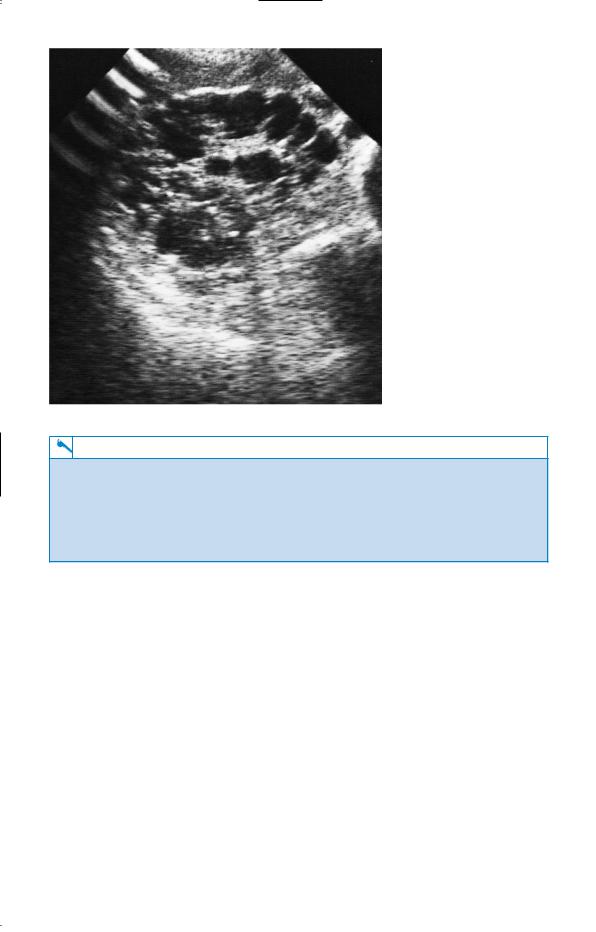
Figure 26.1 Renal ultrasound demonstrating multiple cysts.
KEY POINTS
•Patients with ADPKD are often asymptomatic.
•ADPKD patients may present with loin pain or haematuria.
•ADPKD is the commonest familial cause of renal failure.
•ADPKD is the most likely cause of bilateral renal masses.
•Family members who may have ADPKD should be advised to have their blood pressure measured and a renal ultrasound.
73