Материал: 100_Cases_in_Clinical_Medicine

CASE 41: ABDOMINAL PAIN
History
A 25-year-old woman presents to her general practitioner (GP) complaining of pain in her right loin radiating in to the right iliac fossa. The pain developed 24 h earlier and has become progressively more severe. She has not had her bowels open for 3 days.
She feels alternately hot and cold and is sweaty. She has no pain on passing urine and no urinary frequency. Her last menstrual period was 3 weeks ago. She has had the occasional episode of acute cystitis, approximately on a 2-yearly basis. She has no other significant past medical history.
Examination
She looks unwell and is febrile at 38.5°C. Her pulse rate is 112/min, and blood pressure 104/66 mmHg. Examination of the cardiovascular and respiratory systems is otherwise unremarkable. She is tender to palpation in the right loin and right iliac fossa. There is guarding, and rebound in the right iliac fossa. Bowels sounds are sparse.
INVESTIGATIONS
|
|
Normal |
Haemoglobin |
14.3 g/dL |
11.7–15.7 g/dL |
White cell count |
17.2 % 109/L |
3.5–11.0 % 109/L |
Platelets |
366 % 109/L |
150–440 % 109/L |
Sodium |
139 mmol/L |
135–145 mmol/L |
Potassium |
4.3 mmol/L |
3.5–5.0 mmol/L |
Urea |
10.3 mmol/L |
2.5–6.7 mmol/L |
Creatinine |
96 &mol/L |
70–120 &mol/L |
C-reactive protein (CRP) |
232 mg/L |
!5 mg/L |
Urinalysis: trace protein; trace blood; nitrites negative
Abdominal X-ray: normal
Questions
•What is the diagnosis?
•How would you manage this patient?
109

ANSWER 41
This woman has acute appendicitis. Acute appendicitis classically presents with a short history of central abdominal pain which rapidly localizes to the right iliac fossa. There is guarding and ‘board-like’ rigidity and rebound tenderness in the right iliac fossa. Untreated, some cases will resolve spontaneously, whereas others will perforate leading to localized or generalized peritonitis. Rarely a delayed diagnosis may result in acute appendicitis progressing to an appendix mass consisting of a haemorrhagic oedematous mass in the ileocaecal region.
In this woman the right iliac fossa symptoms and signs together with the raised white cell count and CRP are consistent with appendicitis. The symptoms of loin pain and presence of blood and protein in the urine mimicking a urinary tract infection suggest the appendix may be retrocaecal. Patients with retro-ileal appendicitis often have little abdominal pain, but irritation of the ileum can lead to severe diarrhoea and vomiting. Patients may also present with subacute intestinal obstruction due to intestinal ileus, or urinary retention due to pelvic peritonitis.
!Differential diagnosis of acute appendicitis
•In young adults the differential diagnoses include irritable bowel syndrome, nonspecific mesenteric adenitis, ruptured ectopic pregnancy, twisted or haemorrhagic ovarian cysts, infection of the Fallopian tubes and urinary tract infections.
•In older patients perforated peptic ulcers, diverticulitis, Crohn’s disease and carcinoma of the colon are important differential diagnoses.
The treatment is appendicectomy as soon as urinary tract infection has been excluded. Ultrasound or computed tomography (CT) scanning can be performed if there is uncertainty as to the clinical diagnosis. However a significant number of patients will have a normal appendix removed.
 KEY POINTS
•Retrocaecal appendicitis can mimic acute pyelonephritis.
•In young women ectopic pregnancy and gynaecological pathology can mimic appendicitis.
•Elderly patients may present with atypical symptoms and signs.
110

CASE 42: POSTOPERATIVE DETERIORATION
History
The medical team is asked to review a postoperative surgical patient. A 62-year-old lady had been admitted 10 days previously to have a right hemicolectomy performed for a caecal carcinoma. This was discovered on colonoscopy which was performed to investigate an iron-deficiency anaemia and change in bowel habit. She is otherwise fit with no significant medical history. She is a retired teacher. She neither smokes nor drinks alcohol and is on no medication. Her preoperative serum creatinine was 76 &mol/L. The initial surgery was uneventful, and she was given cefuroxime and metronidazole as routine antibiotic prophylaxis. However the patient developed a prolonged ileus associated with abdominal pain. On postoperative day 5, the patient started to spike fevers up to 38.5°C and was commenced on intravenous gentamicin 80 mg 8 hourly in addition to the other antibiotics. Over the next 5 days the patient remained persistently febrile, with negative blood cultures. In the last 24 h, she has also become relatively hypotensive with her systolic blood pressure being about 95 mmHg despite intravenous colloids. Her urine output is now 15 mL/h.
Examination
She is unwell and sweating profusely. She is jaundiced. Her pulse rate is 110/min regular, blood pressure 95/60 mmHg and jugular venous pressure is not raised. Her heart sounds are normal. Her respiratory rate is 30/min. Her breath sounds are normal. Her abdomen is tender with guarding over the right iliac fossa. Bowel sounds are absent.
INVESTIGATIONS
|
|
Normal |
Haemoglobin |
8.2 g/dL |
11.7–15.7 g/dL |
Mean corpuscular volume (MCV) |
83 fL |
80–99 fL |
White cell count |
26.3 % 109/L |
3.5–11.0 % 109/L |
Platelets |
94 % 109/L |
150–440 % 109/L |
Sodium |
126 mmol/L |
135–145 mmol/L |
Potassium |
5.8 mmol/L |
3.5–5.0 mmol/L |
Bicarbonate |
6 mmol/L |
24–30 mmol/L |
Urea |
36.2 mmol/L |
2.5–6.7 mmol/L |
Creatinine |
523 &mol/L |
70–120 &mol/L |
Glucose |
2.6 mmol/L |
4.0–6.0 mmol/L |
Albumin |
31 g/L |
35–50 g/L |
Bilirubin |
95 mmol/L |
3–17 mmol/L |
Alanine transaminase |
63 IU/L |
5–35 IU/L |
Alkaline phosphatase |
363 IU/L |
30–300 IU/L |
Trough gentamicin level |
4.8 mg/mL |
!2.0 mg/mL |
Urinalysis: ' blood; ' protein; granular casts and epithelial cells
Questions
•What are the causes of this patient’s acute renal failure?
•How would you further investigate and manage this patient?
111

ANSWER 42
This patient has postoperative acute renal failure due to a combination of intra-abdominal sepsis and aminoglycoside nephrotoxicity. Her sepsis is due to an anastomotic leak with a localized peritonitis which has been partially controlled with antibiotics. Her sepsis syndrome is manifested by fever, tachycardia, hypotension, hypoglycaemia, metabolic acidosis (low bicarbonate) and oliguria. The low sodium and high potassium are common in this condition as cell membrane function becomes less effective. The elevated white count is a marker for bacterial infection and the low platelet count is part of the picture of disseminated intravascular coagulation. Jaundice and abnormal liver function tests are common features of intraabdominal sepsis. Aminoglycosides (gentamicin, streptomycin, amikacin) cause auditory and vestibular dysfunction, as well as acute renal failure. Risk factors for aminoglycoside nephrotoxicity are higher doses and duration of treatment, increased age, pre-existing renal insufficiency, hepatic failure and volume depletion. Aminoglycoside nephrotoxicity usually occurs 7–10 days after starting treatment. Monitoring of trough levels is important although an increase in the trough level generally indicates decreased excretion of the drug caused by a fall in the glomerular flow rate. Thus, nephrotoxicity is already established by the time the trough level rises.
This patient needs urgent resuscitation. She requires transfer to the intensive care unit where she will need invasive circulatory monitoring with an arterial line and central venous pressure line to allow accurate assessment of her colloid and inotrope requirements. She also needs urgent renal replacement therapy to correct her acidosis and hyperkalaemia. In a haemodynamically unstable patient like this, continuous haemofiltration is the preferred method. The patient also needs urgent surgical review. The abdomen should be imaged with either ultrasound or computed tomography (CT) scanning to try to identify any collection of pus. Once haemodynamically stable, the patient should have a laparotomy to drain any collection and form a temporary colostomy.
KEY POINTS
•Postoperative acute renal failure is often multifactorial due to hypotension, sepsis and the use of nephrotoxic drugs such as aminoglycosides and non-steroidal anti-inflammatory drugs (NSAIDs).
•Aminoglycoside drugs are extremely valuable for treating Gram-negative infections, but levels must be monitored to avoid toxicity.
•Sepsis syndrome must be recognized early and treated aggressively to reduce the morbidity and mortality of this condition.
112
CASE 43: ABDOMINAL DISCOMFORT
History
A 64-year-old woman is referred to outpatients with a 6-month history of mild abdominal discomfort. This has been intermittent and involved the right iliac fossa mainly. There has been no particular relation to eating or to bowel movements. Over this time her appetite has gone down a little and she thinks that she has lost around 5 kg in weight. The intensity of the pain has become slightly worse over this time and it is now present on most days.
Over the last 6 weeks she has developed some new symptoms. She has developed a different sort of cramping abdominal pain located mainly in the right iliac fossa. This pain has been associated with a feeling of the need to pass her motions and often with some diarrhoea. During these episodes her husband has commented that she looked red in the face but she has associated this with the abdominal discomfort and the embarrassment from the urgent need to have her bowels open.
There is no other relevant previous medical history. She has smoked 15 cigarettes daily for the last 45 years and she drinks around 7 units of alcohol each week. She has noticed a little breathlessness on occasions over the last few months and has heard herself wheeze on several occasions. She has never had any problems with asthma and there is no family history of asthma or other atopic conditions.
She worked as a school secretary for 30 years and has never been involved in a job involving any industrial exposure. She has no pets. She has lived all her life in London and her only trip outside the UK was a day trip to France.
INVESTIGATIONS
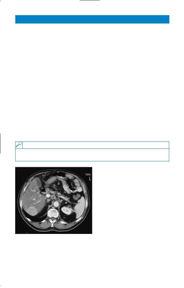
A computed tomography (CT) scan of her abdomen was performed and is shown in Fig. 43.1.
Figure 43.1 Computed tomography scan of the abdomen.
Questions
•What diagnoses should be considered?
•What investigations should be performed?
113