Материал: 100_Cases_in_Clinical_Medicine
CASE 65: SORE THROAT
History
A normally healthy man aged 28 years developed an acute sore throat for which he consulted his general practitioner (GP). A diagnosis of acute pharyngitis was made, presumed streptococcal, and oral penicillin was prescribed. The sore throat gradually improved, but 5 days later the patient noted a rash on his arms, legs and face, and painful ulceration of his lips and mouth. These symptoms rapidly worsened, he felt very unwell and presented to the emergency department. There was no relevant previous medical history or family history. He has had sore throats occasionally in the past but they have settled with throat sweets from the chemist.
Examination
He looked ill and had a temperature of 39.2°C. There were erythematous tender nodules on his arms, legs and face, and ulcers with some necrosis of the lips and buccal and pharyngeal mucosae. The rest of the examination was normal.
INVESTIGATIONS
|
|
Normal |
Haemoglobin |
13.8 g/dL |
13.3–17.7 g/dL |
White cell count |
14.8 % 109/L |
3.9–10.6 % 109/L |
Platelets |
334 % 109/L |
150–440 % 109/L |
Blood film: neutrophil leucocytosis
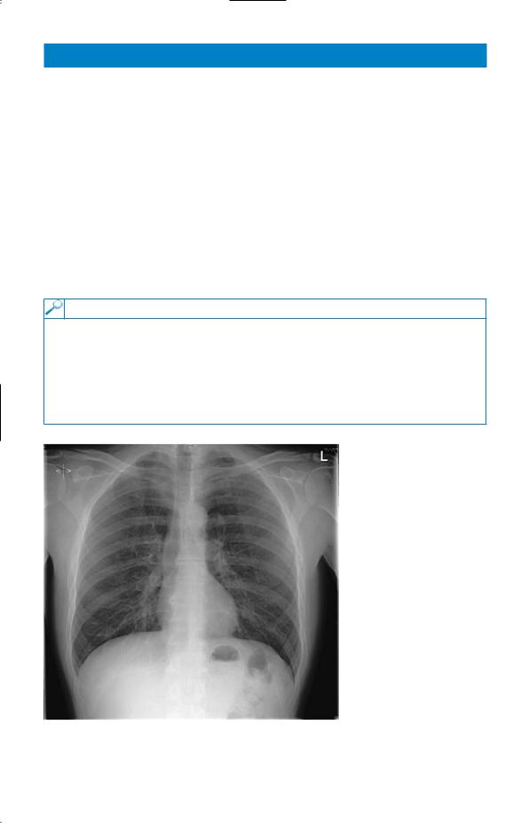
His chest X-ray is shown in Fig. 65.1.
Figure 65.1 Chest X-ray.
Questions
•What is the diagnosis?
•What is the management?
169

ANSWER 65
The diagnosis is acute drug hypersensitivity causing severe erythema multiforme (Stevens–Johnson) syndrome. The pointers to this diagnosis are the rapidity of onset and its timing related to starting the penicillin, antibiotics being the commonest group of drugs causing this syndrome, and the form and distribution of the lesions. The chest X-ray shown is normal.
!Differential diagnoses of the rash
•Streptococcal (presumed) infection spreading to the soft tissues; this is much less common in young healthy patients compared to the elderly; its distribution would be diffuse rather than discrete lesions, and was excluded by negative culture of the lesions.
•Acute leukaemia or neutropenia can present with mucosal ulceration, but not these skin lesions, and these diagnoses are excluded by the blood count and film.
Drugs other than penicillin should be considered as a cause, e.g. analgesics for the original painful throat. The patient had taken a few doses of paracetamol, leaving the penicillin as the likeliest candidate by far as the cause.
!Management
Management consists of:
•stopping the penicillin and substituting an alternative antibiotic if required: cultures were negative in this case at this stage
•a short course of steroids, e.g. 30 mg prednisolone daily for 5 days to reduce the inflammation
•observe for secondary infection of the ulcers
•analgesia
•warn the patient not to take penicillin or related drugs in the future
•record the penicillin allergy clearly in GP and hospital notes.
KEY POINTS
•A drug history is an essential part of every patient’s history.
•Always consider drugs as a cause of complications during a patient’s illness.
•Drug allergies should be recorded prominently in medical notes.
170
CASE 66: URINARY FREQUENCY
History
A 37-year-old man presents to his general practitioner (GP) with a 5-day story of urinary frequency, dysuria and urethral discharge. In the previous 24 h he had become unwell, feeling feverish and with a painful right knee. He works in an international bank and frequently travels to Asia and Australia, from where he had last returned 2 weeks ago. There is no relevant past or family history and he takes no medication.
Examination
He looks unwell, and has a temperature of 38.1°C. His heart rate is 90/min, blood pressure 124/82 mmHg. Otherwise examination of the cardiovascular, respiratory, abdominal and nervous systems is normal. His right knee is swollen, slightly tender, and there is a small effusion with slight limitation of flexion. There is no skin rash and no oral mucosal abnormality. He has a cream-coloured urethral discharge.
INVESTIGATIONS
|
|
Normal |
Haemoglobin |
17.1 g/dL |
13.3–17.7 g/dL |
White cell count |
16.9 % 109/L |
3.9–10.6 % 109/L |
Platelets |
222 % 109/L |
150–440 % 109/L |
Blood film: neutrophil leucocytosis
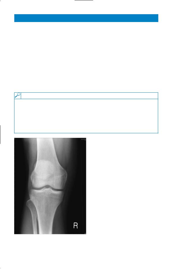
X-ray of right knee is shown in Fig. 66.1.
Figure 66.1 X-ray of the right knee.
Questions
•How would you investigate and manage this patient?
•What is the likely diagnosis?
171

ANSWER 66
The patient has acute gonorrhoea and gonococcal arthritis. The X-ray of the knee is normal. The diagnosis is made by microscopy of the discharge, which should show Grampositive diplococci, and culture of an urethral swab. The swab should be inoculated onto fresh appropriate medium straight away and kept at 37°C until arrival at the laboratory. Immediate treatment on clinical grounds with ciprofloxacin is indicated; penicillin should be reserved for gonorrhoea with known penicillin sensitivity, to prevent the development of resistant strains. Septic monoarthritis is a complication of gonorrhoea; other metastatic infectious complications are skin lesions and, rarely, perihepatitis, bacterial endocarditis and meningitis.
The patient disclosed that he had had unprotected sexual intercourse with prostitutes in Thailand and Singapore; he had had no intercourse following return to the UK so no fol- low-up of contacts was necessary. For advice on precautions and investigation for other sexually transmitted diseases he was referred to the sexually transmitted diseases (STD) clinic.
KEY POINTS
•All students and doctors should be confident in eliciting a sexual history.
•Accurate sexual histories are more likely when the patient feels confidence and empathy with the interviewer.
•Contact tracing is an important element of management of sexually transmitted disease.
172
This page intentionally left blank