Материал: 100_Cases_in_Clinical_Medicine

CASE 67: BACK PAIN
History
A 48-year-old woman presented to her general practitioner (GP) with 3 months’ history of back pain in the mid-thoracic region. The pain was intermittent, worse at night, and relieved by ibuprofen, which she bought herself. She had no other symptoms, and no relevant past or family history. She had never smoked, and drank 10–12 units of alcohol most weeks. She worked part-time stacking the shelves in a supermarket and was a very active and competitive tennis and badminton player.
Examination
She looked well. She indicated that the pain was over the vertebrae of T5/6, but there was no tenderness, swelling or deformity. Her spinal movements were normal.
Her blood pressure was 136/76 mmHg. Cardiovascular, respiratory and abdomen examination were normal.
INVESTIGATIONS
Spinal X-ray was arranged and showed no abnormality. The full blood count, urea creatinine and electrolytes, calcium, alkaline phosphatase and phosphate were all normal, as was urine testing.
She was advised that the pain was musculoskeletal due to exertion at work and sport, and she was prescribed diclofenac for the pain. She was advised to rest from her tennis and badminton.
After a few weeks of improvement, the pain began to get worse, being more severe and occurring for longer periods and seriously disturbing her sleep. She returned to her GP and examination was as before except that there was now some tenderness over her midthoracic spine. The GP arranged another X-ray of the spine (Fig. 67.1).
174
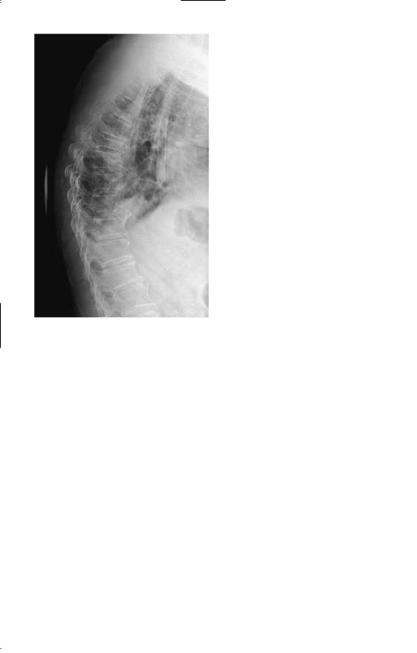
Figure 67.1 Lateral X-ray of the thoracic spine.
Questions
•What is the abnormality in the X-ray?
•What are the likeliest causes?
175

ANSWER 67
The X-ray shows collapse of the T6 vertebra. If there is nothing to suggest osteoporosis or trauma then the commonest cause of this is a tumour metastasis. The tumours that most frequently metastasize to bone are carcinoma of the lung, prostate, thyroid, kidney, and breast. Examination of the patient’s breasts, not done before the X-ray result, revealed a firm mass 1–1.5 cm diameter in the tail of the left breast. Urgent biopsy confirmed a carcinoma and she was referred to an oncologist for further management.
The common lesions affecting the lumbosacral and cervical spine, e.g. inflammation of ligaments and other soft tissues and lesions of the intervertebral discs, are much less common in the thoracic spine, and bony metastases should be considered as a cause of persistent pain in the thoracic spine in patients of an appropriate age.
Review of the first X-ray after the lesion was seen on the second film still failed to identify a lesion, emphasizing the need to repeat an investigation if there is sufficient clinical suspicion of an abnormality, even if an earlier investigation is normal.
Examination of the breasts in women should be part of the routine examination, particularly after the age of 40 years, when carcinoma of the breast becomes common.
KEY POINTS
•Pain in the thoracic vertebrae should raise the possibility of bony metastases in patients over the age of 40 years.
•Repeating previously normal or negative investigations is an important part of a patient’s management when clinical diagnoses remain unconfirmed.
176

CASE 68: A LUMP ON THE SKIN
History
A 66-year-old farmer is referred to a dermatologist for a lesion on his forearm. It is raised, 1.5 cm in diameter, with an irregular margin and a slightly ulcerated centre. It is painless and has appeared over the last 6–8 months. Fifteen years earlier the patient had had a cadaveric renal transplant for renal failure due to chronic glomerulonephritis caused by immunoglobulin A (IgA) nephropathy. This has functioned well, and he has required continuous immunosuppression. Originally this was with prednisolone and azathioprine, but later it was converted to ciclosporin. His only other medication is propranalol for hypertension which he has taken for 20 years. There is no other relevant past or family history. He has never smoked, and drinks 3–6 units of alcohol per week.
Examination
The lesion is as described on the right forearm and there are several solar hyperkeratoses on his cheeks, forehead and scalp (he is bald). The blood pressure is 144/82 mmHg. No other abnormalities are found apart from the transplant kidney in the right iliac fossa.
INVESTIGATIONS
|
|
Normal |
Haemoglobin |
15.4 g/dL |
13.3–17.7 g/dL |
White cell count |
4.6 % 109/L |
3.9–10.6 % 109/L |
Platelets |
356 % 109/L |
150–440 % 109/L |
Sodium |
141 mmol/L |
135–145 mmol/L |
Potassium |
4.2 mmol/L |
3.5–5.0 mmol/L |
Bicarbonate |
29 mmol/L |
24–30 mmol/L |
Urea |
6.7 mmol/L |
2.5–6.7 mmol/L |
Creatinine |
118 &mol/L |
70–120 &mol/L |
Glucose |
5.6 mmol/L |
4.0–6.0 mmol/L |
Urinalysis 'protein; no blood |
|
|
Questions
•What is the likely diagnosis of the lesion on the forearm?
•What factors have contributed to its development?
177

ANSWER 68
The description of the lesion has the characteristic features of a carcinoma of the skin.
The risk factors are his age, the many years exposure to sunlight as farmer, and the chronic immunosuppression. There is an increased risk of several different types of malignancy in patients on chronic immunosuppression, and skin cancer is now well recognized as a frequent complication of chronic immunosuppression unless preventative measures are used. With improving survival rates for transplant patients in general, there is a potential increase in the incidence and prevalence of skin malignancy. Patients on long-term immunosuppression for whatever reason should be strongly advised to avoid direct exposure to sunlight as much as possible, and certainly not to sunbathe, and to use high-factor barrier creams. They should cover their skin in the lighter months (April to September inclusive in the northern hemisphere) – no shorts, sleeveless tops or shirts, and a hat to protect the scalp and forehead. This is particularly irksome but even more important for children and young adults who have a potentially longer period of exposure to sunlight ahead of them. The damage caused to skin by sunlight is cumulative and irreversible, and when transplanted at the age of 50 years this patient had already had over 30 years’ occupational exposure to ultraviolet radiation. His immunosuppression needs to continue and should be kept at as low a dose as is compatible with preventing rejection of his transplant.
The diagnosis of the lesion was made by biopsy, which showed a squamous cell cancer. This was treated by wide excision and skin grafting. An essential part of the follow-up is regular review, at least 6-monthly, of the skin to detect any recurrence, any new lesions or malignant transformation of the solar hyperkeratoses.
KEY POINTS
•Ultraviolet radiation is a cumulative risk factor for skin cancer.
•Preventative measures to reduce exposure to sunlight are an important part of the management of patients on long-term immunosuppression.
178