Материал: 100_Cases_in_Clinical_Medicine

ANSWER 71
The likeliest diagnosis is secondary acute hypoaldosteronism due to failure of the hypo- thalamic-pituitary-adrenal axis caused by the long-term prednisolone. This is a common problem in patients on long-term steroids and arises when there is a need for increased glucocorticoid output, most frequently seen in infections or trauma, including surgery, or when the patient has prolonged vomiting and therefore cannot take the oral steroid effectively. It presents as here with drowsiness and low blood pressure.
The hyponatraemia is another result of the superimposed illness. It is probably due to a combination of reduced intake of sodium owing to the anorexia, and dilution of plasma by the fluid intake. In secondary hypoaldosteronism the renin–angiotensin–aldosterone system is intact and should operate to retain sodium. This is in contrast to acute primary hypoaldosternism (Addisonian crisis) when the mineralocorticoid secretion fails as well as the glucocorticoid secretion, causing hyponatraemia and hyperkalaemia. Acute secondary hypoaldosteronism is often but erroneously called an Addisonian crisis.
Spread of the infection should also be considered, the prime sites being to the brain, with either meningitis or cerebral abscess, or locally to cause a pulmonary abscess or empyema. The patient has a degree of immunosuppression due to her age and the long-term steroid. The dose of steroid is higher than may appear at first sight as the patient is only 50 kg; drug doses are usually quoted for a 70 kg male, which in this case would equate to 10 mg of prednisolone, i.e. an increase of 40 per cent on her dose of 7 mg.
The treatment is immediate empirical intravenous infusion of hydrocortisone and saline. The patient responded and in 5 h her consciousness level was normal and her blood pressure had risen to 136/78 mmHg. Chest X-ray showed bilateral shadowing consistent with pneumonia, but no other abnormality.
KEY POINTS
•Secondary hypoaldosteronism is a medical emergency and requires immediate empirical treatment.
•Patients on long-term steroids should have the dose increased when they have intercurrent illnesses, and replaced systemically when they have persistent vomiting.
184

CASE 72: ABDOMINAL PAIN
History
A 38-year-old woman presents to the emergency department with a 2-h history of severe abdominal pain. The pain is in the right loin and radiates to the right flank and groin and the right side of the vulva. It is colicky and has made her vomit several times. Since the age of 18 years she has had recurrent urinary tract infections, mainly with dysuria and frequency, but she has had at least four episodes of acute pyelonephritis affecting right and left kidneys separately and together. She has not had gross haematuria nor passed stones per urethra. There is no other past history. Her mother had frequent urinary tract infections and died at the age of 61 of a stroke. Over the years the patient has taken irregular intermittent prophylactic antibiotics, but for only approximately a total of 20 per cent of the time. She works in a travelling fairground and has no general practitioner (GP). Access to any previous medical records is not possible as she cannot remember the details of where she was seen or treated. She has had some imaging of the urinary tract but is unsure of the details of the investigations and their results.
Examination
She is ill – flushed and sweating with a pyrexia of 39.2°C. Her heart and chest are normal. She is tender in the right loin. The blood pressure is 150/100 mmHg and funduscopy shows arteriovenous nipping.
INVESTIGATIONS
|
|
Normal |
Haemoglobin |
14.3 g/dL |
11.7–15.7 g/dL |
Mean corpuscular volume (MCV) |
85 fL |
80–99 fL |
White cell count |
18.2 % 109/L |
3.5–11.0 % 109/L |
Platelets |
365 % 109/L |
150–440 % 109/L |
Sodium |
136 mmol/L |
135–145 mmol/L |
Potassium |
5.3 mmol/L |
3.5–5.0 mmol/L |
Bicarbonate |
20 mmol/L |
24–30 mmol/L |
Urea |
16.7 mmol/L |
2.5–6.7 mmol/L |
Creatinine |
384 &mol/L |
70–120 &mol/L |
Urinalysis: ' protein; ''' blood |
|
|
Questions
•What diagnosis would you make?
•How would you interpret the results?
•How would you manage her now and in the long term?
185

ANSWER 72
The pain’s acute onset, colicky nature and radiation are typical of ureteric colic, the likeliest cause of which is a stone. Renal stones can cause infection, or chronic infection can cause scarring which provides a nidus for stone formation.
The high fever and leucocytosis indicate that she has another episode of acute pyelonephritis.
The patient is in renal failure; at this stage it is not clear whether this is all acute, with previous normal renal function, or whether there is underlying chronic renal failure with an acute exacerbation. Both kidneys are affected, as renal function remains normal if one kidney is healthy. Until proved otherwise it must be assumed that any element of acute renal failure is due to obstruction by a stone; her illness is too short for significant prerenal failure due to fluid loss or septicaemia. Acute pyelonephritis per se can cause acute renal failure but this is very uncommon.
She has hypertension. Her blood pressure is raised, but pain and anxiety could easily account for that. However, there is grade I retinopathy.
The overall interpretation at this point is that she is a medical emergency with acute pyelonephritis in an obstructed urinary tract.
The most important investigation now is an ultrasound of the urinary tract. This shows stones in both kidneys; the left kidney is reduced in size to 10 cm, with a scar at its upper pole, and is not obstructed; the right kidney is larger at 11 cm but is obstructed as shown by a dilated renal pelvis and ureter; its true size would be less than 11 cm.
The immediate management is an intravenous antibiotic to treat Gram-negative bacteria, E. coli being the commonest cause of urinary tract infections, after urine and blood samples are taken for culture. Intravenous fluids should be given (she has vomited) according to fluid balance, carefully observing urine output.
The obstruction must be relieved without delay; the method of choice is percutaneous nephrostomy and drainage. In this procedure a catheter is inserted under imaging guidance through the right loin into the obstructed renal pelvis. Not only will this relieve the obstruction but it allows the later injection of X-ray contrast to define the exact site of obstruction (percutaneous nephrostogram). This was done 48 h later and showed hold-up of the contrast at the vesico-ureteric junction, a typical place for a stone to lodge. The patient passed the stone shortly afterwards, as often happens if it is small enough; otherwise it would have to be removed surgically. Her fever, pain and leucocytosis rapidly resolved. Her renal function improved but stabilized at a creatinine of 180 &mol/L, i.e. she has chronic renal failure.
Blood biochemistry revealed no underlying abnormality to cause the stones: calcium, phosphate, alkaline phosphatase and uric acid were normal. The probable cause of her renal disease is reflux nephropathy because of her sex, history of recurrent infections and the scar on the left kidney. There is a familial tendency for this disease, and her mother may have had it. The patient’s children should be screened for it in infancy.
Long-term management comprises prophylactic antibiotics, immediate treatment of acute urinary infections, control of hypertension and regular measurement of renal function. These should be supervised from a fixed base, despite the patient’s peripatetic existence.
KEY POINTS
•An obstructed and infected urinary system is an emergency requiring immediate treatment.
•Prophylactic treatment of recurrent urinary tract infections should be considered in every case, although not necessarily indicated in every one.
186
CASE 73: CHEST PAIN AND SHORTNESS OF BREATH
History
A 25-year-old female accountant complains of shortness of breath, cough and chest pain. The chest pain came on suddenly 6 h previously when she was walking to work. It was a sharp pain in the right side of the chest. The pain was made worse by breathing. It settled over the next few hours but there is still a mild ache in the right side on deep breathing. She felt a little short of breath for the first hour or two after the pain came on but now only feels this on stairs or walking quickly. She has had a dry cough throughout the 6 h.
She smokes 15 cigarettes a day and drinks 10 units of alcohol a week. She uses marijuana occasionally. She is on no medication. Four years ago something very similar happened; she is not sure but thinks that the pain was on the left side of the chest on that occasion. There is no relevant family history.
Examination
She is not distressed or cyanosed. Her pulse is 88/min and blood pressure 128/78 mmHg, respiratory rate is 20/min. Heart sounds are normal. In the respiratory system the trachea and apex beat are not displaced. Expansion seems normal, as is percussion. There is decreased tactile vocal fremitus and the intensity of the breath sounds is reduced over the right side of the chest. There are no added sounds on auscultation.
INVESTIGATIONS
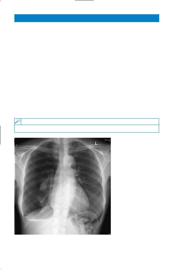
The chest X-ray is shown in Fig. 73.1.
Figure 73.1 Chest X-ray.
Questions
•What does the X-ray show?
•What should be done now?
187

ANSWER 73
The chest X-ray shows a large right pneumothorax. There is a suggestion of a bullous lesion at the apex of the right lung. Pneumothoraces are usually visible on normal inspiratory films but an expiratory film may help when there is doubt. There is no mediastinal displacement on examination or X-ray, movement of the mediastinum away from the side of the pneumothorax would suggest a tension pneumothorax. Although she had symptoms initially, these have settled down as might be expected in a fit patient with no underlying lung disease. A rim of air greater than 2 cm around the lung on the X-ray indicates at least a moderate pneumothorax because of the three-dimensional structure of the lung within the thoracic cage represented on the two-dimensional X-ray.
The differential diagnosis of chest pain in a young woman includes pneumonia and pleurisy, pulmonary embolism and musculoskeletal problems. However, the clinical signs and X-ray leave no doubt about the diagnosis in this woman. Pneumothoraces are more common in tall, thin men, in smokers and in those with underlying lung disease. Further investigations such as computed tomography (CT) scan are not indicated unless there is a suggestion of underlying lung disease.
There is a suggestion that she may have had a similar episode in the past but it may have been on the left side. There is a tendency for recurrence of pneumothoraces, about 20 per cent after one event and 50 per cent after two. Because of this, pleurodesis should be considered after two pneumothoraces or in professional divers or pilots.
The immediate management is to aspirate the pneumothorax through the second intercostal space anteriorly using a cannula of 16 French gauge or more, at least 3 cm long. Small pneumothoraces with no symptoms and no underlying lung disease can be left to absorb spontaneously but this is quite a slow process. Up to 2500 mL can be aspirated at one time, stopping if it becomes difficult to aspirate or the patient coughs excessively. If the aspiration is unsuccessful or the pneumothorax recurs immediately, intercostal drainage to an underwater seal or valve may be indicated. Difficulties at this stage or a persistent air leak may require thoracic surgical intervention. This is considered earlier than it used to be since the adoption of less invasive video-assisted techniques. In this woman the apical bulla was associated with a persistent leak and required surgical intervention through video-assisted minimally invasive surgery.
Marijuana has been reported to be associated with bullous lung disease, and she should be advised to avoid it. Tobacco smoking increases the risk of recurrence of pneumothorax.
KEY POINTS
•The patient should not be allowed to fly for at least 1 week after the pneumothorax has resolved with full expansion of the lung (2 weeks after a traumatic pneumothorax).
•The risk of recurrence will be reduced by stopping smoking.
188