Материал: 41_price2004
resistance to flow is minimized. As the flow rate increased from 5 to 15 íL min-1, the cell-free layer is more apparent. Importantly, the profile of the RBCs flowing through a rectangular channel (Figure 3C and D) in a microchip at flow rates of 2 and 4 íL min-1, respectively, also shows the formation of a cell-free layer. Therefore, even though the channels on a fabricated chip are rectangular, the flow profile is similar to the profile of the RBCs in cylindrical microbore tubing and that seen in vivo. The flow profile of the RBCs through these microchannels, if coupled with more complex designs having channels whose diameters scale such as those in the microcirculation, may result in a vessel mimic that is more realistic than microbore tubing.
The data in Figure 2 showing the amounts of ATP released from RBCs suggest that these cells are subjected to mechanical deformation in the chip channels. Indeed, it has been previously shown that the ATP release from the RBCs is dependent upon the extent of the applied deformation forces.4,5,10,11 However, it is also likely that the duration of that deformation is a determinant of the RBC-derived ATP. In Figure 3E, the amount of ATP derived from RBCs, either mechanically deformed in the microchip channel or fused-silica tubing (50 cm long and 50 ím inside diameter), is shown. These data reveal a slight decrease in ATP release (2.54 ( 0.15 í») from RBCs deformed in a portion of fused-silica tubing when compared to the ATP release (2.59 ( 0.32 íM) from RBCs deformed in the chip channels. However, this amount of RBC-derived ATP is not statistically different from the ATP release from RBCs deformed in the microchip channel having a length of 4.5 cm, a width of 60 ím, and a depth of 38 ím. These data suggest that the diameter of a vessel (or cross sectional area) is more important as a determinant of ATP release from RBCs than the length of the vessel. That is, it appears that the majority of the ATP released from mechanically deformed RBCs occurs when the forces are initially applied, which agrees with previous findings from other groups using filtration as a means of deforming RBCs.4,5 It should also be noted that there was a 13% difference in the cross sectional area of the microchip channel and microbore tubing. However, previous work in this area indicates that such a difference in cross sectional area would not result in significantly different amounts of ATP release from deformed RBCs.10,11 Therefore, not only do the data in 3E imply that similar results can be obtained from both microchip and tubing based vessel mimics, thus providing further evidence that microchip channels may serve as appropriate resistance vessel mimics, but also that vessel length is a minor determinant of ATP release from deformed RBCs.
The data in Figure 4 demonstrate that various hematocrits of RBC samples can be manipulated in the microchip channels. In Figure 4, various RBC hematocrits were prepared from a stock solution of RBCs ( 70% hematocrit after washing the cells in PSS) and pumped through a microchip channel. As shown, the measured amount of RBC-derived ATP increased as the hematocrit increased from 1 (1.53 ( 0.12 íM) to 3.5 (7.62 ( 0.65 íM) to 7% (24.7 ( 0.5 íM). It should also be noted that the discrepancy between RBC-derived ATP for the 7% hematocrit in Figure 4 and Figure 2 is due to RBCs obtained from two separate rabbits on different days. These increasing amounts of ATP release with increments in hematocrit are expected since the number of RBCs available to release ATP is increasing. Moreover, the increased
Figure 4. ATP release from RBCs (that were mechanically deformed by pumping through microchip channels) as a function of increasing the hematocrit from 1 (1.53 ( 0.12 íM) to 7% (24.7 ( 0.50 íM). The error bars represent standard deviations of the set (n ) 3).
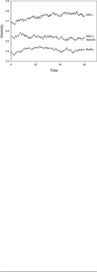
Figure 5. Chemiluminescent intensity from a buffer, a 7% hematocrit of RBCs, and a second aliquot of 7% RBCs stiffened with diamide. The decrease in luminescent intensity (which is directly related to ATP released from the RBCs) for the RBCs incubated with diamide is 62.4%.
number of RBCs in the channel also results in an increase in the mechanical deformation of each RBC since, as shown in Figure 3C and D, the cells migrate to the center of the channel where the resistance to flow is minimized. As the number of cells in the middle of the channel increases, the likelihood of compression by other cells will also increase. Also, although the values for the RBC-derived ATP shown in Figure 4 lie outside of the calibration range, unpublished work in our laboratory has shown that this assay is linear from 0 íM to at least 50 íM.
Finally, to confirm that the measured ATP is indeed due to a mechanical deformation, as opposed to cell lysis, RBCs were incubated with 10 íM diamide for 20 min and pumped through the smaller of the microchip channels described in the Experimental Section. Diamide is an oxidant that is known to stiffen RBC membranes without damaging the cell cytosol.38 Previously, our group and others have shown that ATP release from RBCs correlates with RBC deformability.39,40 That is, if the RBC becomes
(38)Driessen, G. K.; Scheidt-Bleichert, H.; Sobota, A.; Inhoffen, W.; Heidman, H.; Haest, C. M. W.; Kamp, D.; Schmid-Schonbein, H. Pflugers ArcH. 1982, 392, 261-267.
4854 Analytical Chemistry, Vol. 76, No. 16, August 15, 2004

less deformable, as would occur under an oxidant insult, the amount of RBC-derived ATP will decrease. The data in Figure 5 suggest that diamide is indeed stiffening the cell membranes since the amount of ATP released from the RBCs is decreased when incubated with diamide. In Figure 5, the chemiluminescent intensity was measured for a buffer solution containing no ATP or diamide, a 7% hematocrit of RBCs, and another 7% hematocrit of the same RBC sample incubated with the diamide. As shown, the chemiluminescent intensity (after subtracting out the intensity measured for the buffer alone) decreased 62.4%. Importantly, these data confirm that the measured ATP is due to mechanical deformation, since the diamide should have no effect on ATP release from RBCs that are lysed and diamide also has no affect on the chemiluminescence assay for ATP used in this work.
CONCLUSIONS
The ability of channels, patterned in microchips derived from PDMS employing soft lithography methods, to serve as biomimetic arterioles in vivo has been demonstrated. RBCs from rabbits were pumped through channels whose cross sectional dimensions approximate those of resistance vessels in vivo. It has been shown here that the RBCs, when being pumped through the microchip channels, migrate toward the center of the channels, thus leaving a cell-free or skimming layer at the walls of the channel that increases with increments in linear rate of sample through the channels. This type of profile also has been shown to develop in cylindrical tubing and is known to exist in circulatory vessels in vivo.15 Importantly, it has also been demonstrated here that both ATP standards and ATP derived from mechanically deformed
(39)Sprague, R. S.; Stephenson, A. H.; Ellsworth, M. L.; Keller, C.; Lonigro, A. J. Exp. Biol. Med. 2001, 226, 434-439.
(40)Fischer, D. J.; Torrence, N. J.; Sprung, R. J.; Spence, D. M. Analyst 2003, 128, 1163-1168.
RBCs flowing through the microchip channels can be determined “on-chip” via a simple chemiluminscence reaction. Similar to previous findings that used filtration5 or microbore tubing11 to apply forces to the RBCs that these cells would be subjected to in vivo, increasing amounts of ATP are released from the RBCs as the diameter of the vessel mimic (in this case, the microchip channel) decreases. Moreover, the amount of ATP released from RBCs that were deformed in a 50-cm section of microbore tubing was not significantly different from the ATP release from RBCs in a microchannel having a length of 4.5 cm. Thus, it appears that the extent of RBC deformation is more important than the duration of RBC deformation. The work presented here presents a foundation for future work involving microchips channels as resistance vessel mimics. Specifically, microchips will be designed that more closely mimic the complex vascular networking of the vascular beds in the microcirculation of such organs as the lung. Such designs will include branching of the on-chip “vessels” and a uniform scaling of the diameters of these vessels in order to represent the scaling down from arteries to arterioles in vivo.
ACKNOWLEDGMENT
Red blood cells from the research group of Randy Sprague, MD in the Department of Pharmacological and Physiological Sciences at the Saint Louis University School of Medicine are greatly appreciated. The authors thank the research group of Professor Susan M. Lunte (University of Kansas) for use of their cleanroom to fabricate the masters used in this work. This work was supported by the National Institutes of Health (HL 073942-01).
Received for review March 15, 2004. Accepted June 9, 2004.
AC0495992
Analytical Chemistry, Vol. 76, No. 16, August 15, 2004 4855