Материал: BASIC_SURGICAL_TECHNIQUES budapest
Chemical wounds
Acid in a small concentration can irritate the skin or mucous membrane, while a large concentration of it leads to a coagulation necrosis. Treatment: similar to the burn injury. Base leads to the colliquative necrosis. The connective tissue is loosened and the necrosis is extended deeply. Treatment: similar to the burn injury.
Wounds produced by radiation
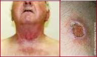
The x-ray (depending to its dose) can lead to erythema and dermatitis. The later complications can be: fibrosis and ulcer (Figure 64.).
Figure 64. Postradiation dermatitis and radiation ulcer
2. Classification of the wounds according to bacterial contamination
Clean wounds (operation or sterile conditions): only the normally present skin bacteria are detectable with no signs of inflammation.
Clean-contaminated wounds: the contamination of clean wounds is endogenous or comes from the environment, the surgical team, or the patient’s skin surrounding the wound. They include opening of the digestive, respiratory or urogenital tract.
Contaminated wounds (significant bacterial contamination): arise when an incision is performed acutely in a non-purulent area or in cases of a leakage from the gastrointestinal tract. Dirty wounds: the contamination comes from an established infection. Examples include: residual nonviable tissues and chronic traumatic wounds.
3. Classification of the wounds depending on the time passed since the trauma Acute (mechanical and other injuries):
-Fresh wound: treatment within 8 h.
-Old wound: ≥8 h after discontinuity of the skin.
Chronic (venous, arterial, diabetic and other ulcers, and skin or soft tissue defects):
-They do not heal within 4 weeks after the beginning of wound management.
-Without treatment, they do not heal within 8 weeks.
4.Classification of the wounds depending on the depth of injury
Grade I: superficial wounds: abrasion; only epidermis and dermis (up to the papillae) are involved.
Grade II: partial-thickness skin wounds: involves the whole thickness of the dermis (intact islands of the hair follicles and sweat glands).
Grade III: full-thickness skin wounds: skin and the subcutaneous tissue are involved (loss of tissue and gaping wound edges).
Grade IV: deep wounds or complex wounds (e.g. lacerations, or vessel and nerve injuries), or wounds of the bone or supporting structures, the opening of body cavities, or penetrating injuries of organs.
51
5.1.2. Management of the accidental wounds
Basic principels
All accidental wounds are considered as infected wounds. There is a need to remove the microorganisms and the nonviable tissues from the wound. An accidental wound shoud be transformed to a surgical wound.
Inspection
Examination of the wound under sterile conditions (cap, mask and gloves).
Anamnesis
-To clarify the circumstances of the injury. When did it happen? The faster we examine the patient, the less possibilty exsits for infection. Is there any accompanying disease which can effect on the healing process (e.g. DM, tumor)? Clarification of the circumstances of the injury can help us to judge about the danger of infection.
-To clarify the state of patient’s vaccination against Tetanus. In the case of infected wound, to give human anti-tetanus Ig. The vaccination and registration are happennig in the admitted traumatological ward.
-Prevention from rabies: in the case of a bite wound (name of vaccine: Rabipur, given at the time of injury and then at the 3th, 7th, 14th, 30th, and 90th days)
Diagnostic procedures
-To exclude the accompanying injuries.
-Examination of the circulation, sensory + motor functions, as well as bone.
Types of the wound management
Temporary wound management (first aid): aim to prevent the secondary infection.
–cleaning of the wound
–hemostasis
–covering
Final primary wound management:
surgical wound closure can be performed if maximum 12 hours is passed since the time of injury.
–cleaning,
–anesthesia,
–excision (< 6–8 h, exception: face, hand),
–sutures (in the case of puncture, bite, shot, and shatterd wounds situating sutures +drain)
Always the primary wound closure is performed in the case of injuries involving the:
–thorasic cavity,
–abdominal wall, and
–the dura matter.
The primary wound closure is contraindicated:
In the following cases, after clearing of the wound and washing it with physiologic saline solution cover it with a sterile bandage and put it in rest. Four to six days later, you can apply the delayed sutures.
–signs of inflammation,
–the wound is strongly contaminated,
–the removal of the foreign body was not successful,
–shattered wounds with blind spaces,
–injuries of persons with especial jobs (e.g. surgeon, butcher, veterinarian, pathologist), and
–bite, shot, and deep punctured wounds.
Need to do: cleaning + covering and after 3-8 days delayed primary wound closure.
52
In the management of the war injuries, we never do the primary wound closure.
–Such these injuries are considered to be infected with aerobic and anaerobic bacteria.
–The readiness of the injured person to fight against the infection is lost.
–Exceptions are: penetrating injuries of the skull, thorax, and abdomen.
Alternatives:
_delayed primary suture (3-8 days)
–to bring the edges of the wound close to each other with the help of adhesive tapes and later to perfom the sutures
–situating sutures + drain
–early secondary wound closure (> 14 days)
–late secondary wound closure (4–6 weeks)
–plastic procedures
Primary delayed suture
If no signs of infection occur within 4–6 days, suturing (or situating suturing) is performed after excision of the wound edges. 3–8 days later: anesthesia + excision (refreshment of the wound edges) and suturing.
Early secondary wound closure
If following the first management of the wound, the excised wound -after inflammation and necrosisstarts to proliferate, then there is a need to refresh the wound edges. 2 weeks after the injury: anesthesia, excision (refreshment of the wound edges), suturing, and draining.
Late secondary wound closure
The proliferating former wound parts and scars should be excised. With greater defects, plastic surgery solutions should also be considered. 4–6 weeks after the injury: anesthesia, excision (of the secondarily healing scar), suturing, and draining.
5.1.3. Surgical wounds
2.1. Determinants of healing of surgical wounds
Preparation of the operating site, hygiene, shaving, disinfection and isolation are. The incision should be parallel to the Langer lines. The skin is stretched, the scalpel is held in a vertical position and the incision is performed until the subcutaneous layer is reached. It is important to be aware of the anatomical structures of the involved area. The muscle is separated along its fascia. The handling of bleeding is of importance.
Skin incision
The skin incision is made on a prepared (cleansed, draped) operative field with taking into consideration of vessels and nerves of the area. During the incision, the surgeon and the assistant stretch the skin. Usually a scalpel is used. The type of the scalpel depends on the site of the incision. The manner of holding the scalpel varies according to the use:
-for a long, straight incision or when there is a need to apply a bigger force, the scalpel is held like a fiddle bow.
-for the delicate, curved incision of fine structures, the scalpel is held like a pen.
53
The important requirements of the skin incision
-The length of the incision should be appropriate for safe surgery.
-Vessels and nerves should not be damaged.
-The skin edges should be smooth.
-The incision is made perpendicularly to the skin with a single definite cut (failed attempts result in ragged edges and prevent wound healing).
-The direction of the incision depends on the location of the organ being operated on.
-The skin is incised parallel to the Langer lines (better wound healing and less scar formation).
-The incision is usually directed toward the operator and from left to right (right-handed person).
-The depth of the incision must be the same throughout the whole length. At the beginning, the tip of the scalpel is inserted perpendicularly into the skin, the cut is made an angle of 45° with the blade of the scalpel (not with the tip!), and the incision is completed with the scalpel held perpendicularly.
-The skin scalpel is discarded into the container after the skin incision. In the deeper layers, another scalpel is used.
Main types of skin incisions based on body region
Neck: Kocher’s transverse incision at the base of the neck (thyroid gland), Thorax: sternotomy, thoracotomy,
Abdomen: subcostal (gallbladder or spleen), median/paramedian laparotomy (this may be upper or lower relative to the umbilicus), transrectal/pararectal/transverse laparotomies, Pfannenstiel suprapubic incision (bladder, uterus or ovaries), McBurney incision (appendectomy), inguinal incisions (hernia).
Closure and dressing of the surgical wounds
Fascia and subcutaneous layer: interrupted stitches. The fat must not be sutured (fat necrosis). Skin: tissue-sparing technique, with accurate approximation of the skin edges. Tension and ischemia of the skin edges are to be avoided. Simple interrupted stitch, Donati vertical mattress suture, Allgöwer stitch, continuous intracutaneous (or subcuticular) suture, Steri-Strips, clips and tissue glues may be applied to close the wound. Dressing: sterile, moist, antibiotic-containnig, non-allergic and non-adhesive dressings. Holding the dressing: adhesive tapes, elastic bandages, and stretchable meshes. The dressing is removed on the 1st postoperative day, and daily in cases of infection.
5.1.4. Process of wound healing, types of it, and its risk factors
Hemostasis–inflammation (days 0-2)
Signs of inflammation (heat, pain, redness, and swelling) are present. The wound fills with blood clot and platelet aggregates, and fibrin production develops. The blood flow is increased, and macrophage and leukocyte mediators are released. Removal of bacterial components.
Granulation–proliferation (days 3-7)
Characterized by the formation of granulation tissue and fibroblasts. Collagen and elastic fibers protect against the infection and they provide a suitable medium for re-epithelization. The healthy sprout is red and does not bleed. Expressed angiogenesis and the connections between the loose extracellular matrix (ECM) and fibronectin are characteristic.
54
Remodeling (lasting for months from day 8)
Maturation = ECM remodeling, and continuous collagen deposition. The scar is characterized by intensive strand formation; the vascularity is reduced and becomes brighter. The ECM is loose and relatively weak (20% of the final strength after 3 weeks). The fibers contract and become smaller and stronger. This contraction can cause a reduction in joint functions. This is pronounced for a year, but remodeling continues for an indefinite time. The final strength of the wound is around70-80% of that of uninjured tissue.
Types of wound healing
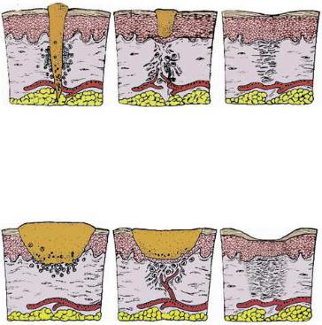
1. The scheme of sanatio per primam intentionem (”p.p. healing”).
According to Galen: “the major aim” of a doctor is the gap-free healing of wounds.
2. The scheme of sanatio per secundam intentionem. The tissue loss is compensated by a granulation tissue “according to the second potential goal of the doctor”. Due to the abacterial or purulent inflammation, the wound is filled with connective tissue which transforms into scar tissue.
Factors influencing wound repair
Some factors infuence the process of wound healing. Among drugs: glucocorticoids inhibit fibroblast activity, protein synthesis and immune responses. Some antibiotics inhibit collagen biosynthesis. Cytostatic agents slow down metabolic processes. Anti-inflammatory agents reduce hyperemia and the blood supply to the wound. General condition, nutrition, protein level, vitamins B, C and K, and trace elements (Zn and Mg) (malnutrition slows down the healing process). Diabetes mellitus: There is a risk of infection, dysfunctioning of the microand macrocirculation, and hyperglycemia can lead to the developement of chronic wounds. Icterus (the accompanying liver dysfunction influences the wound healing); similarly anemia, tumorous conditions and bacterial (or other) infections can influence the porcess of wound healing.
55