Материал: BASIC_SURGICAL_TECHNIQUES budapest
5.1.5. Complications of wound healing
Early complications of wound healing
Seroma: The wound cavity is filled with serous fluid, lymph or blood. Signs: fluctuation, swelling, redness, tenderness and subfebrility. Treatment: sterile puncture ad compression; if repeated, then use a suction drain. It is common after the breast operations.
Hematoma: Due to an inefficient control of bleeding, a short drainage time or anticoagulation therapy. The risk of infection is high. Signs: swelling, fluctuation, pain and redness. Treatment: in the early phase, sterile puncture; later, surgical exploration is required.
Wound disruption: The major types are: partial, superficial (dehiscence), and complete separation (disruption). First, the deeper layers are involved and finally the skin. Local causes: a surgical error (e.g. suturing the fascia with a continuious suture), increased intraabdominal pressure and wound infection. Treatment: in the operating room and under general anesthesia, we apply the U-shaped en masse sutures to relieve tension.
Superficial wound infection
1.A diffuse and superficially spreading inflammation located below the skin (e.g. erysipelas, lymphangitis which is caused by hemolytic streptococci). Treatment: resting position, antibiotic, and dermatological consultation.
2.Localized (circumscribed) infection (e.g. an abscess). It can happen anywhere (e.g. under the skin, between the muscles, subfascially, in the thorax; brain; or liver). Treatment: surgical exploration and drainage. There is always a need to think of a foreign body (corpus alienum, filum suppuratio); it can develop even years later (X- ray examination is always necessary!).
Deep wound infections
1.Diffuse infection (e.g. an anaerobic necrosis). Treatment: surgical exploration, open therapy, rinsing the wound with H2O2, and antibiotics.
2.Localized infection (e.g. empyema), inside the tissues or body cavities (e.g. pleural and joint cavities). Treatment: surgical exploration and drainage (Staphylococcus aureus!).
Mixed wound infection
1.Gangrene: necrotic tissues, putrid and anaerobic infection; a severe clinical picture. Treatment: aggresive surgical debridement and effective and specified (antibiotic) therapy.
2.Generalized reaction: bacteremia, pyaemia, and sepsis.
Prevention of the wound infection
Taking into consideration the general basic rules for handling and treating a wound, a thorough examination, preparation, taking care of asepsis, rapid decision and if needed an extended exploration, to apply atraumatic techniques, and correct handling of the bleeding.
Signs and treatment of the wound infection
Local signs: rubor, tumor, calor, dolor and functio laesa. General signs: a rapid sedimentation rate of the RBCs, leukocytosis, fever, shivering, depression.
General therapy: rest and steam bandage if necessary. In the event of aggravation of the symptoms, wound exploration is performed under local anesthesia, with surgical removal of pus, necrotic tissues or foreign material (tissue sample is taken for bacteriological examination), daily rinsing with 3% H2O2 solution (or with antiseptics, povidone-iodine: Betadine, Braunol), open wound management and daily wound toilette.
56
Late complications of the wound closure
Scar formation in the penetration channels, hyperrophic scar, keloid formation, necrosis, inflammatory infiltration, abscesses and foreign body-containnig absecesses.
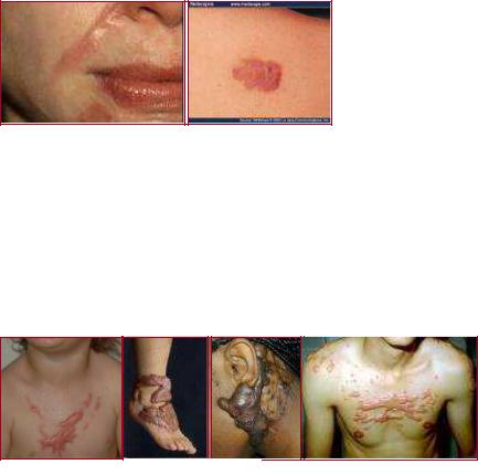
Hypertrophic scar
These develop in areas of thick chorium. They are composed of non-hyalinic collagen fibers and fibroblasts. They are confined to the incision line. Treatment: they regress spontaneously, starting 3-6 months after surgery, and fall back to the level of the skin in 1–2 years (Figure 65.).
Figure 65. Hypertrophic scars
Keloid
These are of unknown etiology, they affect mostly African and Asian populations. They have well-defined edges, with pinkish-brown, emerging, tough structures, which result from the overproliferation of collagen fibers in the subcutaneous tissue of the skin. They particularly affect scars on the presternal and deltoid areas and ears (Figure 66.).They are characterized with subjective complains (e.g. pain, itching sensation and aesthetic problems) and constant development. Treatment: intralesional corticosteroid+local anesthetic injections, postoperative radiation therapy. Prevention: with application of an atraumatic surgical technique.
Figure 66. Keloids
5.2 Bleeding and hemostasis
5.2.1 Hemostasis
This is a natural, life-saving defense mechanism which has three main components: 1. vascular (vasoconstriction), 2. platelet, and 3. clotting. All of these inhibit or decrease bleeding from the vessels. During injury (e.g. a surgical incision), the endothelial damage exposes matrix proteins and collagen. Following this, the platelets clump and adhere to connective tissue at the cut site (adhesion). The platelets then release adenosine diphosphate (ADP), epinephrine, thromboxane A2, and serotonin (release). The binding sites for fibrinogen appear on the platelet membrane and fibrinogen becomes involved in platelet-platelet adhesion (aggregation). ADP and thrombin cause further platelet activation. In this way, the primary thrombus is formed. The clotting cascade is
57
also activated and with catalytic action of thrombin fibrin is produced from fibrinogen. Loss of the circulating blood is termed hemorrage. This can be acute or chronic, primary or secondary. The causes of a secondary hemorrhage can be: infected wounds, inadequate primary wound care, inadequate or traumatic dressings, or necrosis of the vessel wall (e.g. compression, drain, etc.).
Anatomical bleeding
It is originating from a cut or injured bigger vessel. Arterial bleeding is bright red and pulsating with the cardiac function. The volume loss depends on the size of the artery. Venous bleeding is often a continuous flow of dark red blood with lower intensity. The amount of the lost blood and the danger of bleeding are more. If large veins are injured, there will be a possibilty for the air embolism!
Diffuse bleeding
Origin: oozing from denuded or cut surfaces. The continuous loss of blood from oozing can become serious if it remains uncontrolled. Capillary bleeding: a tamponade with dry or wet (warm saline) towels is used to stop oozing. It is improtant to apply a continuous pressure because wiping the wound can remove the alreadyformed thromuses from the end of the capillaries. Parenchymal bleeding: absorbable sutures or gelatin are used. Minor bleeding during skin incision can be controlled by compression of the skin edges with towels.
Classification of bleeding
The patient destiny is determined by the volume of the lost blood and time passed since the bleeding was started. The severity of the bleeding depends on the volume of lost blood/time ratio. The value of this ratio depends on the size of the injured vessel, blood pressure, and the resistances of the surrounding tissues. The clinical outcomes are: bleeding to death, hemorrhagic shock, functional disturbancies due to compression (cardiac tamponade, cerebral hemorrhage, breathlessness), anemia, and so on. To assess hemorrhage, the patient’s mean blood volume must be known (males have ≈ 70 ml/kg (7% of the body weight), while females have ≈ 65 ml/kg. We classify the bleeding based on its severity.
|
Class I |
Class II |
Class III |
Class IV |
Blood loss[ml] |
0–750 |
750–1500 |
1500–2000 |
>2000 |
Mean blood |
15 % |
15–30 % |
30–40 % |
40–50 % |
volume [%] |
|
|
|
|
HR |
< 100 |
> 100 |
> 120 |
> 140 |
MAP |
Normal |
Normal |
Decreased |
Decreased |
RR |
14–20 |
20–30 |
30–35 |
> 35 |
Capillary refill |
Normal |
Slight delay |
> 2 sec |
No filling |
Skin |
Pink, cold |
Pale, cold |
Pale, cold, moist |
Mottled |
Urine |
> 30 ml/h |
20–30 ml/h |
5–15 ml/h |
< 5 ml/h |
Behavior |
Slight anxiety |
Mild anxiety |
Anxious, confused |
Lethargic, confused |
Fluid therapy |
No |
Crystalloid, |
Crystalloid, colloid, |
Crystalloid, colloid, |
|
fluid/crystalloid |
colloid |
blood |
blood |
5.2.2. Direction of hemorrhage
58
Clinically, bleeding can be external (e.g. trauma, or a surgical incision resulting in visible hemorrhage) or internal (e.g. urinary tract: hematuria, respiratory tract: hemoptoa, GIT: hematochezia or melena). The latter can be directed toward body cavities (intracranial hemorrhage, hemothorax, hemascos, hemopericardium, and hemarthros), or among tissues (e.g.hematoma and suffusion). Bleeding can be classified according to the time of surgical interventions: it can be preoperative, intraoperative or postoperative.
Preoperative hemorrhage
Bleeding outside the hospital (see traumatology and anesthesiology). Prehospital care for hemorrhagic injuries includes: maintenance of the airways; ventilation and circulation, the control of an accessible hemorrhage with bandages; direct pressure and tourniquets (these methods have not changed greatly during 2000 years), and the treatment of possible shock with i.v. fluids.
Intraoperative hemorrhage
This can be anatomical and diffuse. Risk factors include drugs used in clotting disorders to reduce clotting (anticoagulants, antiplatelet drugs and thrombolytics), cirrhosis and liver dysfunction (clotting factors defficiency), uremia, hereditary coagulation disorders and sepsis. The main factors influencing intraoperative blood loss:
1.The attitude of the surgeon, his/her training and experience
2.Planning of surgery - selecting the simplest technique-
3.The optimal size of the surgical team. Meticulous attention to bleeding points - skillness of surgeon + proper use of diathermy, laser devices, tissue glues, and minimal invasive techniques-
4.Posture - the level of the operative site should be a little above the level of the heart (e.g. the Trendelenburg position for lower limb, pelvic and abdominal procedures; and the reverse Trendelenburg position for head and neck surgery)-
5.The size of the bleeding vessels.
6.The pressure in the vessels.
7.Hemostasis: the diameter of bleeding vessels decreases spontaneously due to vasoconstriction (more pronounced in arterioles than in venules).
8. To handle the bleeding from arterioles isr ea(“surgical”)ie than that from the diffuse veins Anesthesia (!): Intraoperative bleeding depends much more on the B.P. rather than on the CO; the BP can be maintained at an optimally low level by the anesthesiologist. Various anesthetic techniques are applied to minimize preoperative blood loss:
- If adequate planes of anesthesia and analgesia are ensured, hypertension and tachycardia due to sympathetic overactivity can be avoided
- Controlled anesthesia (increase in intratracheal pressure increase in CVP increase in PCO2increase in B.P.)
- Regional anesthesia (epidural or spinal): where appropriate can leads to 45% reduction in blood loss (sympathicolysis leads to a lower MAP, and spontaneous breathing to a lower CVP)
- With use of the controlled hypotension
- With proper drug therapy of the hypertensive patients
Postoperative bleeding
Causes: Ineffective local hemostasis, a complication of blood transfusion, a previously undetected hemostatic defect, consumptive coagulopathy, or fibrinolysis (a prostate, pancreas or liver operation). Causes of postoperative bleeding starting immediately after the operation:
59
-an unligated bleeding vessel;
-a hematologic problem arising as a result of the operation. Therapy
-If the circulation is unstable, immediate reoperation is essential!
-Action to be taken if the circulation is stable:
-reassessment of the history and medication given,
-checking the body temperature; if it is low, the patient should be warmed,
-laboratory coagulation tests.
5.2.3. Local and general signs and symptoms of bleeding
Local:
Visible signs: hematoma, suffusion, ecchymosis
−Compression[(e.g. breathlessness (pleural cavity, neck)]
−Constriction/compression:
−Cardiac insufficiency (pericardium)
−Increase in ICP (skull)
−compartment syndrome (between muscles)
−Functional disturbancies: hyperperistalsis (GIT bleeding), intestinal paralysis (retroperitoneal hematoma)
General signs: pale skin and mucous membrane, cyanosis, decreased B.P. and tachycardia, difficulty in breathing, sweeting, body temprature is decreased, unconciousness, cardiac and respiratory standstill, laboratory disorders, and signs and symptoms of shock (see later).
5.3. Surgical hemostasis
The aim of local hemostasis is to prevent the flow of blood from the incised or transected vessels. Bleeding which appears in the surgical territory makes the orientation difficult. It is one of the most dangerous complications of the surgery and the biggest obstecle to wound healing. These give the reasons to perform a proper intraoperative hemostasis. Methods are: 1. mechanical, 2. thermal, or 3. chemical.
5.3.1. Mechanical methods – temporary and final interventions
Digital pressure
When possible, direct pressure is combined with elevation of the bleeding site above the level of the heart. Applied over a proximal arterial pressure point. Intraoperative maneuvers [e.g. the Pringle (Báron) maneuver: compression of the vessels at the porta hepatis, in Hungary it was first applied by Sándor Báron in 1910]
Tourniquet
There is no completely safe tourniquet duration. In most cases, a tourniquet can be left in place for 2 hours without causing permanent nerve or muscle damage. A tourniquet is commonly used in hand surgery to produce a bloodless operative field.
Ligation
Artery forceps (Péan, Kocher, mosquito, etc.): this is the most commonly used method of hemostasis in surgery. The source of the bleeding should be grasped by a hemostat with minimal
60