Материал: BASIC_SURGICAL_TECHNIQUES budapest
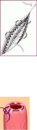
Intracutaneus continuous suture
This runs in intacutan plane parallel to the skin surface; it enters the skin at the beginning and comes out at the end. It produces a fine scar. At both ends, the thread can be tied or taped to the skin (Figure 54.).
Figure 54. Intracutaneus continuous suture
Purse-string suture
The openings of the gastrointestinal tract (e.g. in appendectomy) are closed with this suture. An atraumatic needle and thread are used. It is a suture for a circular opening, running continuously around it. The wound edges are then inverted into the opening with dressing forceps and the threads are pulled and knotted (Figure 55.).
Figure 55. Purse-sting suture
4.3.3. Removing sutures
The time of removal (usually within 3–14 days) depends on the location of the suture (sutures are removed later from a field which is under tension), the blood supply of the operative field (sutures can be removed earlier from an area that has good circulation) and the general condition of the patient. Sutures on the face can be removed after 3–5 days, those on the skin of the head and the abdominal wall after 7–10 days, those on the trunk and the joints after 10–14 days, those on the hand and arm after 10 days, and those on the leg and foot after 8–14 days.
Removing simple interrupted sutures
After careful disinfectioning of the wound, the suture is grasped and gently lifted up with a thumb forceps. The thread should be cut as close to the skin as possible so that no thread which was outside the skin should be pulled through the wound. In this way, infection of the wound can be avoided.
Removing continous sutures
In the case of locked continuous sutures, the thread is cut between the knot -which is located at
46
one endand skin and then the thread is removed. In continuous subcuticular sutures, one end of the suture is cut above the skin and the other end is pulled out in the direction of the wound.
Removing wound clips
Done with the Michel clip applicator and remover. The ring of one end of the clip is grasped with a tissue forceps, the edge of the remover is placed between the clip and the wound line, beneath the apex of the clip. The instrument is closed, the clip will open and the teeth of the clip will come out of the skin.
47
5. WOUNDS AND THE BASIC RULES OF HANDLING THEM BLEEDING,
HEMOSTASIS,
THE PROCESS OF WOUND HEALING
5.1. Wounds and the basic rules of their handling
Wound is a circumscribed injury which is due to an external force and can involve any tissue or organ. It can be mild, severe, or even lethal. As a result of wound, the liquid and element parts of the blood are lost and the protective function of the skin is disturbed. These result the microorganisms and the forighn bodies to enter into the body. The exposure of body cavities and internal organs means a further risk.
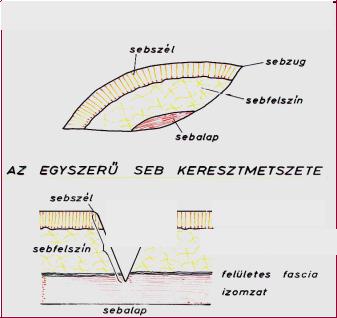
In simple wounds, skin, mucous membrane, subcutaneous tissue, superficial fascia, and the muscles (partially) can be injured (Figure 56.). This needs a simple wound management which can be done even by a nonspecialist. In the case of compound wounds beside those tissues which can be injured in a simple wound, there are injuries of muscles, tendons, vessels, nerves or bones. The joint space may become opened and if the body cavities are injured then there will be a possibility for injury of the internal organs as well. Management of such these wounds is done by a specialist and there is need for well-equipped institutes and a work team (consisting of surgeon, traumatologist, and anaesthesiologist).
The accidental wounds can be either open or closed. Wounds can result from mechanical, thermal or chemical forces and irradiation. The surgical wounds are the sign of the surgical incisions or interventions. They are usually produced in sterile circumstances and during a surgical intervention the surgeon close them layer by layer.
Parts of wound
Wound edge |
|
|
Wound |
|
|
|
corner |
|
|
|
|
|
|
Surface of |
|
|
|
the wound |
|
|
|
|
|
|
|
Base of the wound |
|
|
|
|
|
|
Cross section of a simple wound |
||
Wound edge |
|
|
|
|
|
|
|
|
Wound |
Skin surface |
|
cavity
Surface of |
|
Subcutaneus tissue |
|||
|
|
|
|
||
the wound |
|
|
|
|
|
|
Superficial fascia |
||||
|
|
|
|
||
|
|
|
|
||
|
|
|
|
Muscle layer |
|
|
|
|
|
|
|
|
Base of the wound |
|
|
||
|
|
|
|
||
|
|
|
|
|
|
Figure 56. Simple wound
48
5.1.1. Classification of the accidental wounds
1. Classification based on the origion of the wound Mechanical wounds
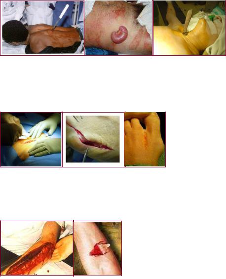
Punctured wound (vulnus punctum) is caused by a sharp pointed tool. It is misleading and sometimes seems to be negligible. Dangers: possibilty for an anaerobic infection, there can be a possibilty for injury of big vessels and nerves which are located deep to the wound (Figure 57.).
Figure 57. Punctured wounds
Incised wound (vulnus scissum): is caused by sharp objects; sharp wound edges are extending up to the base of the wound; the angles of the wound are narrow. All tissues are cut sharply and without any shattering. All surgical incisions belong to this type. It exhibits the best healing (Figure 58.).
Figure 58. Incised wounds
Cut wound (vulnus caesum): is similar to an incised wound, but a blunt additional force also plays a role in its appearance. The degree of shattering is big in the cut tissues and the edges of the wound are uneven (Figure 59.).
Figure 59. Cut wounds
Crush wound (vulnus contusum): is caused by a blunt force and can be either open or closed. The essence is: there is a pressure injury between the external force and the hard (bony) base. The edges are uneven and torn. The bleeding is negligible, but the pain is proportionately greater than would be expected from the size of the injury (termed wound stupor)(Figure 60.).
49
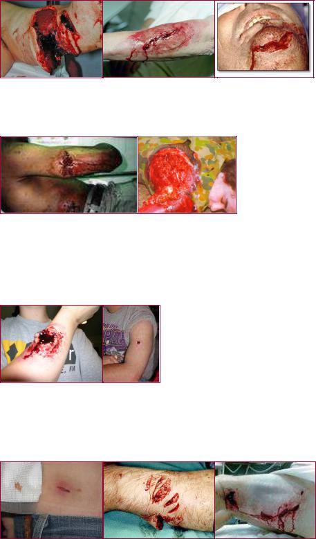
Figure 60. Crush wounds
Torn wound (vulnus lacerum): is caused by great tearing or pulling forces and can result in the incomplete amputation of certain body parts (Figure 61.).
Figure 61. Torn wounds
Shot wound (vulnus sclopetarium): consists of an aperture, a slot tunnel and a possible output. A shot from a close distance is usually accompanied by some degree of burn injury at the aperture. Characteristic features are the incorporated foreign materials (e.g. textile fibers and bullets) and the altered injuries of the tissues locating in the course of the solt tunnel (Figure 62.).
Figure 62. Shot Wouds
Bite wound (vulnus morsum) is a ragged wound with crushed tissue characterized by the shape of the biting teeth and the force of the bite. It is also accompanied by the features of torn wounds. There is a high risk of infection.It is produced by either animals or humans (Figure 63.).
Figure 63. Bite wounds
50