Материал: Презентация экг инглиш
Внимание! Если размещение файла нарушает Ваши авторские права, то обязательно сообщите нам
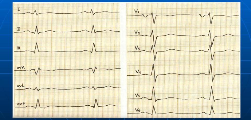
Left atrium hypertrophy
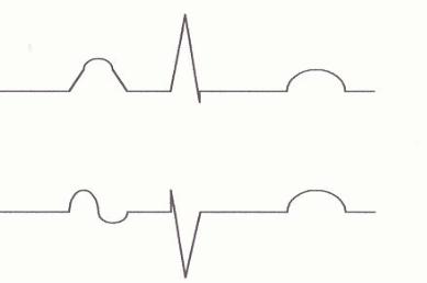
Right atrium hypertrophy is detected by ―P pulmonale‖: high (>2.5 mm) acute Р in II, III, AVF and right chest leads (V1- 2).
Right atrium hypertrophy is typical in chronic pulmonary diseases (pulmonary heart) and tricuspid valves incompetence.
II, III, AVF
V1-2
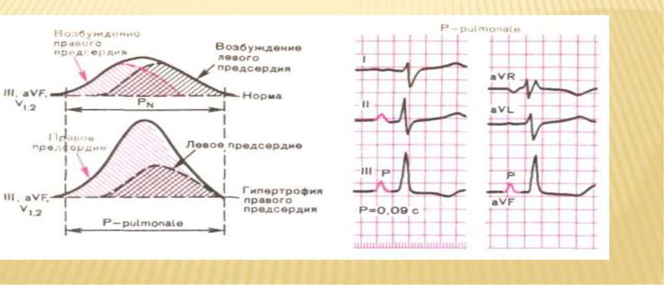
Right atrium hypertrophy

ECG in left bundle branch block is characterized: - the QRS interval is 0,12 sec in duration;
-wide and deformed QRS has the form of qR in I, II, V5- 6; rS in III,
aVF, V1-2 (the shape of the ventricular complexes resembles that of right
ventricular extrasystoles);
-disconcordance of ST, T and the main wave of QRS; - levogram.
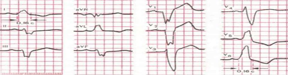
left bundle branch block
Источник: https://studfile.net/preview/16448279/