Материал: 100_Cases_in_Clinical_Medicine

drugs (NSAIDs) for the pain and he may require bed rest. If his disease relapses he should be referred to a rheumatologist. He and his wife should be referred to the sexually transmitted disease clinic for counselling and testing for other sexually transmitted diseases such as hepatitis B, HIV and syphilis.
KEY POINTS
•The most likely causes of an acute large joint monoarthritis are a septic arthritis and a seronegative arthritis.
•Septic arthritis must be recognized and treated as a medical emergency.
39

CASE 16: PAIN IN THE KNEE
History
An 80-year-old woman presents to her general practitioner (GP) with pain and swelling in her left knee. The pain began 2 days previously and she says that the knee is now hot, swollen and painful on movement. In the past she has a history of mild osteoarthritis of the hips. She has occasional heartburn and indigestion. She had a health check 6 months previously and was told that everything was fine except for some elevation of her blood pressure which was 172/102 mmHg and her creatinine level, which was around the upper limit of normal. The blood pressure was checked several times over the next 4 weeks and found to be persistently elevated and she was started on treatment with 2.5 mg bendrofluamethizide. The last blood pressure reading was 138/84 mmHg. There is no relevant family history. She has never smoked and her alcohol consumption averages four units per week. She takes occasional paracetamol for hip pain.
Examination
Her blood pressure is 142/86 mmHg. The temperature is 37.5°C and the pulse 88/min. There is grade 2 hypertensive retinopathy. There is no other abnormality on cardiovascular or respiratory examination. In the hands there are Heberden’s nodes over the distal interphalangeal joints.
The left knee is hot and swollen with evidence of effusion in the joint with a positive patellar tap. There is pain on flexion beyond 90 degrees. The right knee appears normal.
INVESTIGATIONS
|
|
Normal |
Haemoglobin |
12.1 g/dL |
11.7–15.7 g/dL |
White cell count |
12.4 % 109/L |
3.5–11.0 % 109/L |
Platelets |
384 % 109/L |
150–440 % 109/L |
Erythrocyte sedimentation rate (ESR) |
48 mm/h |
!10 mm/h |
Sodium |
136 mmol/L |
135–145 mmol/L |
Potassium |
3.6 mmol/L |
3.5–5.0 mmol/L |
Urea |
7.3 mmol/L |
2.5–6.7 mmol/L |
Creatinine |
116 &mol/L |
70–120 &mol/L |
Glucose |
10.8 mmol/L |
4.0–6.0 mmol/L |
An X-ray of the knees is performed and the result is shown in Fig. 16.1.
40
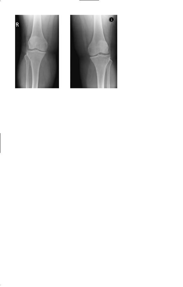
Figure 16.1 X-ray of both knees.
Questions
•What is the likely diagnosis?
•What is the appropriate management?
41

ANSWER 16
The clinical picture is one of acute monoarthritis. The patient has a history of some hip pains but this and the Heberden’s nodes are common findings in an 80-year-old woman, related to osteoarthritis. The blood results show a raised white cell count and ESR, a raised blood sugar, and renal function at the upper limit of normal.
!Differential diagnoses of pain in the knee
The differential diagnosis includes trauma, septic arthritis, gout and pseudogout.
The recent introduction of a thiazide diuretic for treatment of the hypertension increases the suspicion of gout. Pseudogout is caused by deposition of calcium pyrophosphate crystals and would be expected to show calcification in the articular cartilage in the knee joint. The X-rays here show some joint space narrowing but no calcification in the articular cartilage. The fever, high white cell count and ESR are compatible with acute gout. The raised glucose may also be a side-effect of thiazide diuretics. If this remains after the acute arthritis has subsided then it may need further treatment. Precipitation of gout by thiazides is more likely in older women, particularly in the presence of renal impairment and diabetes. It may involve the hands, be polyarticular and can affect existing Heberden’s nodes.
The serum uric acid level is likely to be raised, but this occurs commonly without evidence of acute gout. The definitive investigation is aspiration of the joint. The fluid should be sent for culture and inspection for crystals. A high white cell count would be expected in an acute inflammatory arthritis. The diagnosis is made from the needle-like crystals of uric acid which are negatively birefringent under polarized light, unlike the positively birefringent crystals of calcium pyrophosphate.
In this case the pain in the joint was partly relieved by the aspiration. Treatment with a non-steroidal anti-inflammatory drug should be covered by a proton pump inhibitor in view of her history of heartburn and indigestion. The thiazide diuretic was changed to an angiotensin-converting enzyme inhibitor as treatment for her hypertension, and the blood glucose settled.
KEY POINTS
•A careful drug history is an essential part of the history.
•Thiazide diuretics can precipitate diabetes and gout, especially in the elderly.
42

HAEMATOLOGY
CASE 17: EASY BRUISING
History
A 36-year-old woman presents to her general practitioner (GP) complaining of spontaneous bruising mainly on her legs. The bruising has been noticeable over the last 4–6 weeks. She cannot remember any episodes of trauma. In addition her last two menstrual periods have been abnormally heavy, and she has suffered a major nosebleed. She otherwise feels well, and is working full time as a secretary. There is no significant past medical history. She is married with one daughter aged 11 years. There is no family history of a bleeding disorder. She is a non-smoker and drinks a small amount of alcohol socially.
Examination
On examination there are multiple areas of purpura on her legs and to a lesser extent on her abdomen and arms. The purpuric lesions vary in colour from black–purple to yellow. There are no signs of anaemia, but there are two bullae in the mouth and there is spontaneous bleeding from the gums. There are no retinal haemorrhages on funduscopy. Blood pressure is 118/72 mmHg. Examination of the cardiovascular, respiratory and abdominal systems is unremarkable.
INVESTIGATIONS
|
|
Normal |
Haemoglobin |
10.9 g/dL |
11.7–15.7g/dL |
Mean corpuscular volume (MCV) |
83 fL |
80–99 fL |
White cell count |
4.3 % 109/L |
3.5–11.0 % 109/L |
Platelets |
4 % 109/L |
150–440 % 109/L |
Sodium |
139 mmol/L |
135–145 mmol/L |
Potassium |
4.3 mmol/L |
3.5–5.0 mmol/L |
Urea |
5.4 mmol/L |
2.5–6.7 mmol/L |
Creatinine |
76 &mol/L |
70–120 &mol/L |
Glucose |
4.3 mmol/L |
4.0–6.0 mmol/L |
Clotting screen: normal |
|
|
Blood film: decreased platelets |
|
|
Questions
•What is the likely diagnosis?
•How would you further investigate and manage this patient?
43