Материал: 100_Cases_in_Clinical_Medicine

ANSWER 34
This patient has a number of important symptoms, particularly the generalized seizures, auditory hallucinations, fever, arthralgia and alopecia. Investigations show low haemoglobin, white cells and platelets with impaired renal function and blood, protein and cells in the urine. The CSF contains white cells and a high protein content but no organisms. This is a multisystem disease and the symptoms and investigations are explained best by a diagnosis of systemic lupus erythematosus (SLE). SLE is an autoimmune condition which is about nine times more common in women than men, and is especially common in African-Caribbean and Asian individuals. It varies in severity from a mild illness causing a rash or joint pains, to a life-threatening multisystem illness. In the brain, SLE causes a small-vessel vasculitis and can present with depression, a schizophrenia-like psychosis, fits, chorea and focal cerebral/spinal cord infarction. Lumbar puncture usually shows a raised leucocyte count and protein level. A Coombs’-positive haemolytic anaemia may occur. Leucopenia and thrombocytopenia are common. Glomerulonephritis is another common manifestation of lupus and may present with microscopic haematuria/proteinuria, nephrotic syndrome or renal failure. Arthritis commonly affects the proximal interphalangeal and metacarpophalangeal joints and wrists, usually as arthralgia without any deformity.
!Differential diagnosis of the combination of headaches/psychiatric features/fits
•Meningitis/encephalitis
•‘Recreational’ drug abuse, e.g. cocaine
•Cerebral tumour
•Acute alcohol withdrawal: delirium tremens
•Hypertensive encephalopathy
This patient needs urgent antihypertensive treatment to lower her blood pressure, and anticonvulsant treatment. Blood should be sent for anti-DNA antibodies (present in SLE) and complement C3 and C4 levels (depressed in SLE). A renal biopsy will provide histological evidence of the severity of the lupus nephritis. As soon as active infection has been excluded, treatment should be started with intravenous steroids and cytotoxic agents such as cyclophosphamide. Plasma exchange may be added in severe or resistant cases.
KEY POINTS
•SLE is particularly common in young African-Caribbean women.
•SLE may present with predominantly neurological or psychiatric features.
•A low white cell count or low platelet numbers are often a suggestive feature of SLE.
94

CASE 35: SWELLING IN THE NECK
History
A 38-year-old man presents to his general practitioner (GP) complaining of a painless lump on the right side of his neck. This has been present for about 2 months and seems to be enlarging. He has had no recent throat infections. He has been feeling generally unwell and has lost about 5 kg in weight. The patient has also developed drenching night sweats. Simultaneously he has noticed severe generalized itching. He has had no significant past medical history. He is an accountant, and married with three children. He neither smokes nor drinks alcohol and is not taking any regular medication.
Examination
His temperature is 37.8°C. There is a smooth, firm 3 % 4 cm palpable mass in the right supraclavicular fossa. There are also lymph nodes 1–2 cm in diameter, palpable in both axillae and inguinal areas. His oropharynx appears normal. There are multiple excoriations of his skin. His pulse rate is 100/min regular and blood pressure 112/66 mmHg. Examination of his cardiovascular and respiratory systems is normal. On abdominal examination, there is a mass palpable 3 cm below the left costal margin. The mass is dull to percussion and it is impossible to palpate its upper edge. Neurological examination is normal.
INVESTIGATIONS
|
|
Normal |
Haemoglobin |
11.6 g/dL |
13.3–17.7 g/dL |
Mean corpuscular volume (MCV) |
87 fL |
80–99 fL |
White cell count |
12.2 % 109/L |
3.9–10.6 % 109/L |
Platelets |
321 % 109/L |
150–440 % 109/L |
Erythrocyte sedimentation rate |
74 mm/h |
!10 mm/h |
Sodium |
138 mmol/L |
135–145 mmol/L |
Potassium |
4.2 mmol/L |
3.5–5.0 mmol/L |
Urea |
5.2 mmol/L |
2.5–6.7 mmol/L |
Creatinine |
114 &mol/L |
70–120 &mol/L |
Calcium |
2.44 mmol/L |
2.12–2.65 mmol/L |
Phosphate |
1.1 mmol/L |
0.8–1.45 mmol/L |
Total protein |
65 g/L |
60–80 g/L |
Albumin |
41 g/L |
35–50 g/L |
Bilirubin |
16 mmol/L |
3–17 mmol/L |
Alanine transaminase |
22 IU/L |
5–35 IU/L |
Alkaline phosphatase |
228 IU/L |
30–300 IU/L |
Urinalysis: no protein; no blood |
|
|
Questions
•What is the likely diagnosis?
•How would you investigate and manage this patient?
95

ANSWER 35
Transient small nodes in the neck or groin are common benign findings. However, a 3 % 4 cm mass of nodes for 2 months is undoubtedly abnormal. Persistent lymphadenopathy and constitutional symptoms suggest a likely diagnosis of lymphoma or chronic leukaemia. Sarcoidosis and tuberculosis are possible but less likely diagnoses. Lymph nodes are normally barely palpable, if at all. The character of enlarged lymph nodes is very important. In acute infections the nodes are tender and the overlying skin may be red. Carcinomatous nodes are usually very hard, fixed and irregular. The nodes of chronic leukaemias and lymphomas are non-tender, firm and rubbery. The distribution of enlarged lymph nodes may be diagnostic. Repeated minor trauma and infection may cause enlargement of the locally draining lymph nodes. Enlargement of the left supraclavicular nodes may be due to metastatic spread from bronchial and nasopharyngeal carcinomas or from gastric carcinomas (Virchow’s node). However, when there is generalized lymphadenopathy with or without splenomegaly, a systemic illness is most likely. The typical systemic symptoms of lymphoma are malaise, fever, night sweats, pruritus, weight loss, anorexia and fatigue. Fever indicates extensive disease, and may be associated with night sweats. Severe skin itching is a feature of some cases of lymphoma and other myeloproliferative illnesses.
The incidence of lymphoma is greatly increased in patients who are immunosuppressed, such as organ transplant recipients and patients with HIV infection.
!Major differential diagnosis of generalized lymphadenopathy
•Infections: infectious mononucleosis or ‘glandular fever’ (caused by Epstein–Barr virus infection), toxoplasmosis, cytomegalovirus infection, acute HIV infection, tuberculosis, brucellosis and syphilis.
•Inflammatory conditions: systemic lupus erythematosus, rheumatoid arthritis and sarcoidosis.
•Lymphomas or chronic lymphocytic leukaemia.
The most likely clinical diagnosis in this man is lymphoma. The patient should be referred to a local haemato-oncology unit. He should have a lymph-node biopsy to reach a histological diagnosis, and a computed tomography (CT) scan of the thorax, abdomen and bone marrow to stage the disease. CT scanning is a non-invasive and effective method of imaging retroperitoneal, iliac and mesenteric nodes. Positron-emission tomography (PET) combined with CT increases the sensitivity for detecting disease (Fig. 35.1), and is useful for assessing response to treatment. The patient will require treatment with radiotherapy and chemotherapy. Radiotherapy alone is reserved for patients with limited disease, but this patient has widespread disease. He should be given allopurinol prior to starting chemotherapy, to prevent massive release of uric acid as a consequence of tumour lysis, which can cause acute renal failure.
96
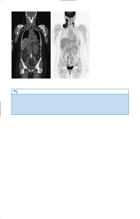
Figure 35.1 CT–PET image showing increased activity in enlarged lymph nodes, particularly in the right side of the neck.
KEY POINTS
•The character and distribution of abnormal lymph nodes is helpful in reaching a diagnosis.
•Lymphadenopathy affecting two or more separate groups of nodes suggests lymphoma or a systemic infection.
•CT–PET scanning allows accurate staging of disease and assessment of maintenance of remission in response to treatment.
97
CASE 36: ABDOMINAL PAIN
History
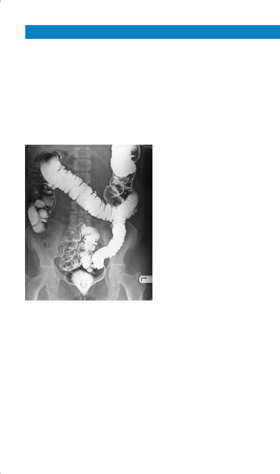
A 74-year-old woman has a 10-year history of intermittent lower abdominal pain. The pain has been colicky in nature and is associated with a feeling of distension in the left iliac fossa. It is generally relieved by passing flatus or faeces. She tends to be constipated and passes small pieces of faeces. Four years previously she passed some blood with her bowel motion and had a barium enema performed. This is shown in Fig. 36.1. Over the last week her pain has worsened and now she has continuous pain in the left iliac fossa and feels generally unwell. Her appetite has been poor over this same time. She has not had her bowels open over the last 2 days. In her previous medical history she had a hysterectomy for fibroids 20 years ago. There is a family history of ischaemic heart disease and diabetes mellitus. She lives alone and does her own cooking and shopping.
Figure 36.1 Barium enema.
Examination
She has a temperature of 38.5°C and is tender with a vague impression of a mass in the left iliac fossa. There is no guarding or rebound tenderness and the bowel sounds are normal. Her pulse is 84/min and blood pressure is 154/88 mmHg. There are no abnormalities to find in the respiratory system.
98