Материал: 100_Cases_in_Clinical_Medicine

ANSWER 30
This picture of loss of menstruation (secondary amenorrhoea), weight loss (to a body mass index (BMI) of 14.0) and hypokalaemic, hypochloraemic metabolic alkalosis fits well with a diagnosis of anorexia nervosa. This is a disorder usually of teenagers or young adults characterized by severe weight loss, a disorder of body image (the patient perceiving themself as being fat despite being objectively thin) and amenorrhoea (or, in men loss of libido or potency). It is commoner in women than men. Often sufferers from this condition work in a profession where personal image is very important, e.g. models, actresses, ballet dancers, and there may be a trigger of an emotional upset such as break-up of a relationship or failure in important examinations. Patients may abuse purgatives or diuretics or cause selfinduced vomiting. Some patients exhibit the bulimic behaviour of recurrent bouts of overeating and self-induced vomiting. Patients often deny that they are ill or that they need medical attention. There is marked wasting with obvious bony prominences. The skin is dry with growth of lanugo hair over the neck, cheeks and limbs as in this woman. There is usually a sinus bradycardia and hypotension. Severe physical complications include proximal myopathy, cardiomyopathy and peripheral neuropathy.
!Major causes of secondary amenorrhoea
•Hypothalmic/pituitary pathology, e.g. hypopituitarism, hyperprolactinaemia.
•Gonadal failure, e.g. autoimmune ovarian failure, polycystic ovaries.
•Adrenal disease, e.g. Cushing’s disease.
•Thyroid disorders, e.g. both hypothyroidism and hyperthyroidism.
•Severe chronic illnesses, e.g. cancer, chronic renal failure.
A number of interrelated mechanisms cause the metabolic alkalosis in this patient. The vomiting causes a net loss of hydrogen and chloride ions, causing alkalosis and hypochloraemia. The loss of fluid by vomiting leads to a contracted plasma volume with consequent secondary hyperaldosteronism to conserve sodium and water, but with renal loss of potassium, due to its secretion in preference to sodium and the fact that fewer hydrogen ions are available for secretion by the renal tubules. These events combine to give the typical picture of an alkalosis with low chloride and raised bicarbonate in the blood, and urine which contains excess potassium and very little chloride. Measurement of 24-h urinary chloride excretion is helpful. A low urinary chloride excretion (!10 mmol/day) implies vomiting, whereas higher values suggest diuretic abuse.
This patient should be referred to a unit with a special interest in eating disorders. Other serious physical illnesses should be excluded with the appropriate investigations. Plasma levels of luteinizing hormone (LH), follicle-stimulating hormone (FSH) and oestrogens will be low. Often such patients are admitted for several weeks in an attempt to make them gain weight. This involves a high-calorie diet with support from the medical and nursing team. Supportive psychotherapy tackles the patient’s disordered perception of their body image.
KEY POINTS
•Anorexia nervosa is a common cause of amenorrhoea in young women.
•Hypokalaemic metabolic alkalosis is the characteristic metabolic abnormality.
•Anorexia nervosa may be associated with abuse of diuretics or purgatives.
84
This page intentionally left blank

CASE 31: PAIN IN THE BACK
History
A 75-year-old woman presents to her general practitioner (GP) complaining of severe back pain. This developed suddenly a week previously after carrying a heavy suitcase at the airport. The pain is persistent and in her lower back. She has had increasing problems with back pain over the past 10 years, and her family have commented on how stooped her posture has become. Her height has reduced by 10 cm over this period. Her past medical history is notable for severe chronic asthma. She takes courses of oral corticosteroids, and use steroid inhalers on a regular basis. She fell 2 years ago and sustained a Colles’ fracture to her left wrist. She developed the menopause at age 42 years. She smokes 30 cigarettes a day, and drinks four bottles of wine a week.
Examination
She has a thoracic kyphosis. She is tender over the L4 vertebra. She has a moon-face, abdominal striae and a number of bruises on her arms and thighs. She is not anaemic, and examination is otherwise unremarkable.
INVESTIGATIONS
|
|
Normal |
Haemoglobin |
11.9 g/dL |
11.7–15.7 g/dL |
Mean corpuscular volume (MCV) |
105 fL |
80–99 fL |
White cell count |
6.2 % 109/L |
3.5–11.0 % 109/L |
Platelets |
358 % 109/L |
150–440 % 109/L |
Erythrocyte sedimentation rate (ESR) |
8 mm/h |
!10 mm/h |
Sodium |
143 mmol/L |
135–145 mmol/L |
Potassium |
4.9 mmol/L |
3.5–5.0 mmol/L |
Urea |
5.9 mmol/L |
2.5–6.7 mmol/L |
Creatinine |
102 &mol/L |
70–120 &mol/L |
Calcium |
2.42 mmol/L |
2.12–2.65 mmol/L |
Phosphate |
1.26 mmol/L |
0.8–1.45 mmol/L |
Alkaline phosphatase |
156 IU/L |
30–300 IU/L |
X-ray of the lumbar spine is shown in Fig. 31.1. |
|
|
86
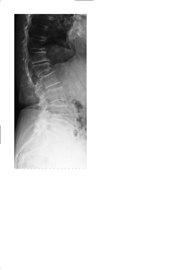
Figure 31.1 X-ray of the lumbar spine.
Questions
•What is the likely diagnosis?
•How would you manage this patient?
87

ANSWER 31
This woman has vertebral collapse due to osteoporosis. The symptoms of osteoporosis are deformity, localized pain and fracture. The loss of height is typical, and is usually noted more by others than the patient. The back pain is due to collapse of the vertebrae. This can occur spontaneously or in association with a recognized stress such as carrying a heavy load. Examination confirms loss of trunk height, thoracic kyphosis and proximity of the ribs to the iliac crest.
!The differential diagnoses of osteoporosis
•Multiple myeloma
•Metastatic carcinoma, particularly from the prostate, breast, bronchus, thyroid and kidney
•Osteomalacia
•Hyperparathyroidism
•Steroid therapy or Cushing’s syndrome
This patient has several risk factors for osteoporosis. Firstly she is aged 75 years, and ageing is associated with bone loss. Secondly she has been postmenopausal for over 30 years. Premenopausal ovarian production of oestrogens help to preserve bone mass. Thirdly she has been on oral and inhaled corticosteroids for her asthma for years. Finally, excess alcohol intake may also be a factor. Her red cells are macrocytic, which is consistent with heavy alcohol intake. Alcohol can lead to an increased incidence of falls and fractures. She has no clinical evidence of thyrotoxicosis or hypopituitarism which can cause osteoporosis.
This woman should have blood tests to exclude myeloma, cancer and metabolic bone disease. Patients with myeloma are anaemic with a raised ESR and a monoclonal paraprotein on serum protein electrophoresis. In contrast to metabolic bone diseases biochemical measurements (serum calcium, alkaline phosphatase and parathormone (PTH)) in osteoporosis are normal. She should have plain X-rays of her spine. Collapse of the vertebral body will manifest as irregular anterior wedging affecting some vertebrae and not others (L1 and L4). A dual-energy X-ray absorptiometry (DEXA) scan can be performed to assess the severity of the osteoporosis, but treatment is indicated anyway with a fracture at this age.
She should have her dose of corticosteroids reduced to the minimum required to control her asthmatic symptoms, using the inhaled routes as far as possible. She should be started on calcium and vitamin D supplements and a bisphosphonate to try to reduce her bone loss. Oestrogen-based hormone replacement therapy is only used for symptoms associated with the menopause because of the increased incidence of thromboembolism and endometrial carcinoma. Newer treatments for osteoporosis include strontium and parathyroid hormone.
KEY POINTS
•Osteoporosis is common in the elderly.
•Bone loss is more rapid in women than men.
•DEXA scan is the method of choice of screening for osteoporosis.
•There are increasingly effective treatments available for the treatment of osteoporosis.
88