Материал: 100_Cases_in_Clinical_Medicine

ABBREVIATIONS
AAT |
alanine aminotransferase |
ACE |
angiotensin-converting enzyme |
ACTH |
adrenocorticotrophic hormone |
ADH |
antidiuretic hormone |
ADPKD |
autosomal dominant polycystic kidney disease |
APTT |
activated partial thromboplastin time |
ARAS |
atherosclerotic renal artery stenosis |
AVP |
arginine vasopressin |
BCG |
bacille Calmette–Guérin |
BMI |
body mass index |
CJD |
Creutzfeld–Jakob disease |
CMV |
cytomegalovirus |
COPD |
chronic obstructive pulmonary disease |
CRP |
C-reactive protein |
CSF |
cerebrospinal fluid |
CT |
computed tomography |
CVP |
central venous pressure |
DDAVP |
L-deamino-8-D-arginine vasopressin |
DEXA |
dual-energy X-ray absorptiometry |
DOT |
directly observed therapy |
DVT |
deep vein thrombosis |
EBV |
Epstein–Barr virus |
ECG |
electrocardiogram |
EEG |
electroencephalogram |
EMG |
electromyogram |
ERCP |
endoscopic retrograde cholangiopancreatography |
ESR |
erythrocyte sedimentation rate |
FER |
forced expiratory ratio |
FEV1 |
forced expiratory volume in 1 s |
FMD |
fibromuscular dysplasia |
FSH |
follicle-stimulating hormone |
FVC |
forced vital capacity |
GnRH |
gonadotrophin-releasing hormone |
GP |
general practitioner |
HbA1c |
haemoglobin A1c |
HDL |
high-density lipoprotein |
5-HIAA |
5-hydroxyindole acetic acid |
5-HT |
5-hydroxytryptamine |
IBS |
irritable bowel syndrome |
ICU |
intensive care unit |
IgG |
immunoglobulin G |
IgM |
immunoglobulin M |

INR |
international normalized ratio |
IPF |
idiopathic pulmonary fibrosis |
ITP |
idiopathic thrombocytopenic purpura |
JVP |
jugular venous pressure |
LDL |
low-density lipoprotein |
LH |
luteinizing hormone |
MCV |
mean corpuscular volume |
MRSA |
methicillin-resistant Staphylococcus aureus |
NAD |
nothing abnormal detected |
NGU |
non-gonococcal urethritis |
NSAID |
non-steroidal anti-inflammatory drug |
NSIP |
non-specific interstitial pneumonitis |
nvCJD |
new-variant CJD |
paCO2 |
arterial partial pressure of carbon dioxide |
pCO2 |
partial pressure of carbon dioxide |
PEF |
peak expiratory flow |
PET |
positron-emission tomography |
pO2 |
partial pressure of oxygen |
SIADH |
syndrome of inappropriate ADH secretion |
SLE |
systemic lupus erythematosus |
STD |
sexually transmitted diseases |
TIA |
transient ischaemic attack |
TIBC |
total iron-binding capacity |
TNF |
tissue necrosis factor |
TSH |
thyroid-stimulating hormone |
T4 |
thyroxine |
TTP |
thrombotic thrombocytopenic purpura |
UIP |
usual interstitial pneumonia |
VDRL |
venereal disease research laboratory |
VLDL |
very low-density lipoprotein |
WOSCOPS |
West of Scotland Coronary Prevention Study |

Section 1 SYSTEMS-RELATED CASES
This page intentionally left blank
CARDIOLOGY
CASE 1: DIZZINESS
History
A 75-year-old man is brought to hospital with an episode of dizziness. He still feels unwell when he is seen 30 min after the onset. He was well until the last 6 months, since when he has had some falls, irregularly. On some occasions he lost consciousness and is unsure how long he has been unconscious. On a few occasions he has fallen, grazing his knees, and on others he has felt dizzy and has had to sit down but has not lost consciousness. These episodes usually happened on exertion, but once or twice they have occurred while sitting down. He recovers over 10–15 min after each episode.
He lives alone and most of the episodes have not been witnessed. Once his granddaughter was with him when he blacked out. Worried, she called an ambulance. He looked so pale and still that she thought that he had died. He was taken to hospital, by which time he had recovered completely and was discharged and told that he had a normal electrocardiogram (ECG) and chest X-ray.
There is no history of chest pain or palpitations. He has had gout and some urinary frequency. A diagnosis of benign prostatic hypertrophy has been made for which he is on no treatment. He takes ibuprofen occasionally for the gout. He stopped smoking 5 years ago. He drinks 5–10 units of alcohol weekly. The dizziness and blackouts have not been associated with alcohol. There is no relevant family history. He used to work as an electrician.
Examination
He is pale with a blood pressure of 96/64 mmHg. The pulse rate is 33/min, regular. There are no heart murmurs. The jugular venous pressure is raised 3 cm with occasional rises. There is no leg oedema; the peripheral pulses are palpable except for the left dorsalis pedis. The respiratory system is normal.
INVESTIGATIONS
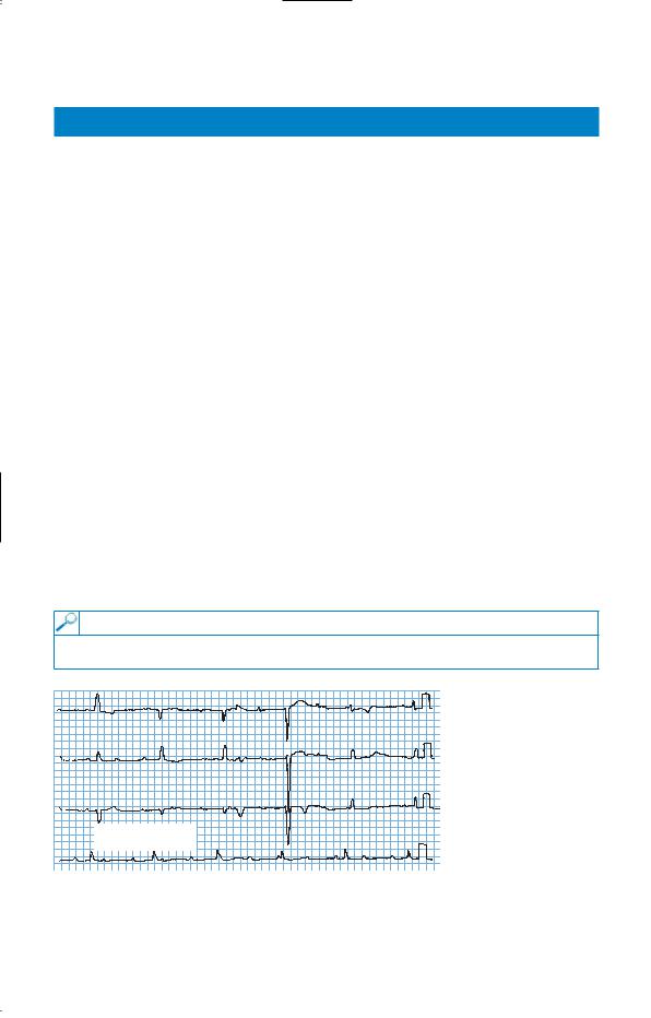
• The patient’s ECG is shown in Fig. 1.1.
|
aVR |
V1 |
V4 |
II |
aVL |
V2 |
V5 |
III |
aVF |
V3 |
V6 |
Rhythm strip:II |
|
|
|
25 mm/s; 1 cm/mV |
|
|
|
Figure 1.1
Questions
•What is the cause of his blackout?
•What does the ECG show?
3