Материал: 100_Cases_in_Clinical_Medicine
ANSWER 1
The blackouts do not seem to have had any relationship to posture. They have been a mixture of dizziness and loss of consciousness. The one witnessed episode seems to have been associated with loss of colour. This suggests a loss of cardiac output usually associated with an arrhythmia. This may be the case despite the absence of any other cardiac symptoms. There may be an obvious flushing of the skin as cardiac output and blood flow return.
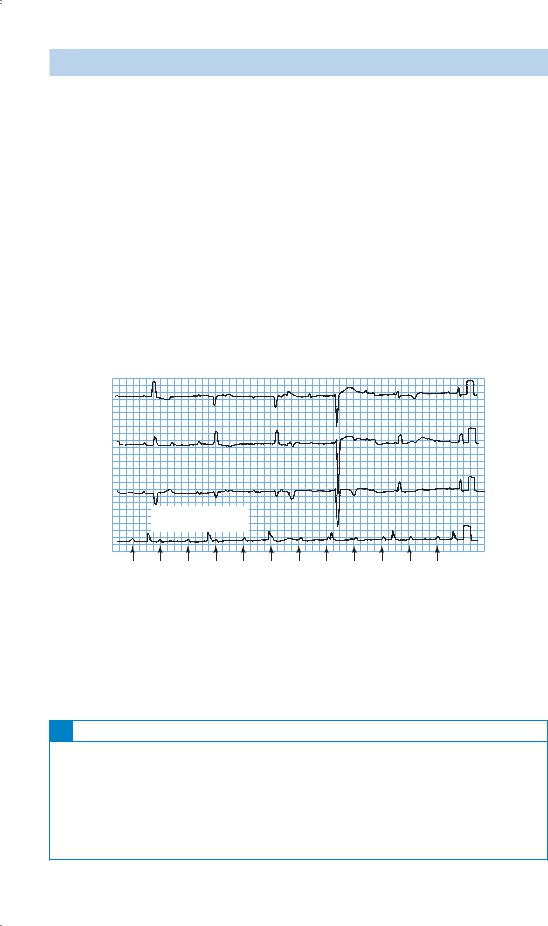
The normal ECG and chest X-ray when he attended hospital after an episode do not rule out an intermittent conduction problem. On this occasion the symptoms have remained in a more minor form. The ECG shows third-degree or complete heart block. There is complete dissociation of the atrial rate and the ventricular rate which is 33/min. The episodes of loss of consciousness are called Stokes–Adams attacks and are caused by self-limited rapid tachyarrhythmias at the onset of heart block or transient asystole. Although these have been intermittent in the past he is now in stable complete heart block and, if this continues, the slow ventricular rate will be associated with reduced cardiac output which may cause fatigue, dizziness on exertion or heart failure. Intermittent failure of the escape rhythm may cause syncope.
|
aVR |
V1 |
V4 |
II |
aVL |
V2 |
V5 |
III |
aVF |
V3 |
V6 |
Rhythm strip:II |
|
|
|
25 mm/s; 1 cm/mV |
|
|
|
Figure 1.1 Electrocardiogram showing complete heart block, p-waves arrowed.
On examination, the occasional rises in the jugular venous pressure are intermittent ‘cannon’ a-waves as the right atrium contracts against a closed tricuspid valve. In addition, the intensity of the first heart sound will vary.
!Differential diagnosis
The differential diagnosis of transient loss of consciousness splits into neurological and vascular causes. A witness is very helpful in differentiation. Neurological causes are various forms of epilepsy, often with associated features. Vascular causes are related to local or general reduction in cerebral blood flow. Local reduction may occur in transient ischaemic attacks or vertebrobasilar insufficiency. A more global reduction, often with pallor, occurs with arrhythmias, postural hypotension and vasovagal faints.
4

The treatment should be insertion of a pacemaker. If the rhythm in complete heart block is stable then a permanent pacemaker should be inserted as soon as this can be arranged. This should be a dual-chamber system pacing the atria then the ventricles (DDD, dual sensing and pacing, triggered by atrial sensing, inhibited by ventricular sensing) or possibly a ventricular pacing system (VVI, pacing the ventricle, inhibited by ventricular sensing). If there is doubt about the ventricular escape rhythm then a temporary pacemaker should be inserted immediately.
KEY POINTS
•When a patient suffers transient loss of consciousness, a careful history from a witness may help with the diagnosis.
•Normal examination and ECG do not rule out intermittent serious arrhythmias.
•Large waves in the jugular venous pressure are usually regular giant v-waves in tricuspid regurgitation or intermittent cannon a-waves in complete heart block.
5

CASE 2: CHEST PAIN
History
A 34-year-old male accountant comes to the emergency department with acute chest pain. There is a previous history of occasional stabbing chest pain for 2 years. The current pain had come on 4 h earlier at 8 pm and has been persistent since then. It is central in position, with some radiation to both sides of the chest. It is not associated with shortness of breath or palpitations. The pain is relieved by sitting up and leaning forward. Two paracetamol tablets taken earlier at 9 pm did not make any difference to the pain.
The previous chest pain had been occasional, lasting a second or two at a time and with no particular precipitating factors. It has usually been on the left side of the chest although the position had varied.
Two weeks previously he had an upper respiratory tract infection which lasted 4 days. This consisted of a sore throat, blocked nose, sneezing and a cough. His wife and two children were ill at the same time with similar symptoms but have been well since then. He has a history of migraine. In the family history his father had a myocardial infarction at the age of 51 years and was found to have a marginally high cholesterol level. His mother and two sisters, aged 36 and 38 years, are well. After his father’s infarct he had his lipids measured; the cholesterol was 5.1 mmol/L (desirable range !5.5 mmol/L). He is a non-smoker who drinks 15 units of alcohol per week.
Examination
His pulse rate is 75/min, blood pressure 124/78 mmHg. His temperature is 37.8°C. There is nothing abnormal to find in the cardiovascular and respiratory systems.
INVESTIGATIONS
•A chest X-ray is normal. The haemoglobin and white cell count are normal. The creatine kinase level is slightly raised. Other biochemical tests are normal.
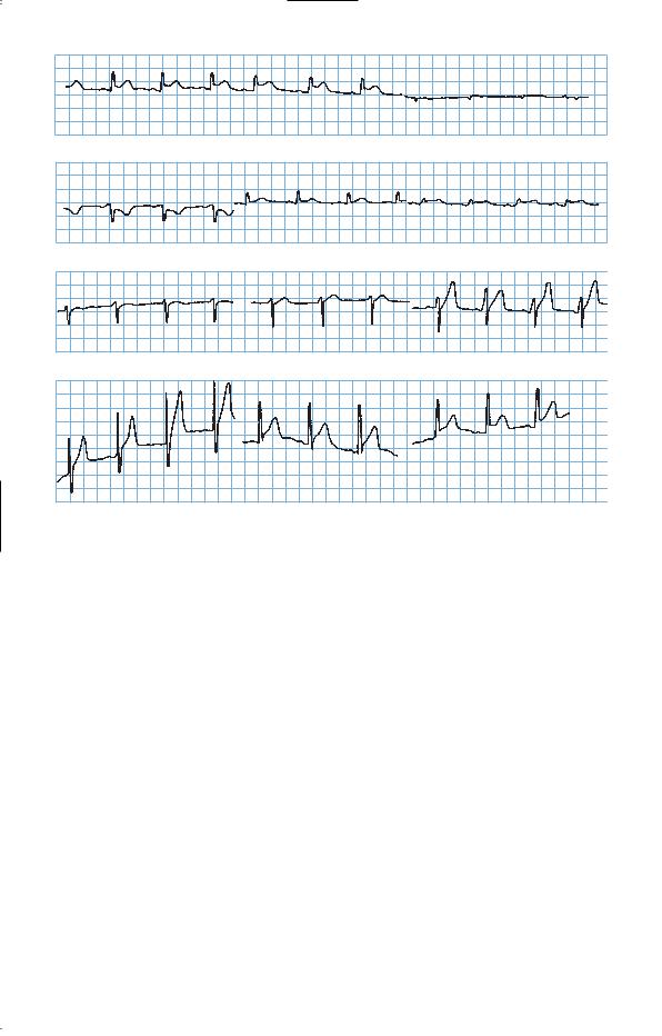
•The ECG is shown in Fig. 2.1.
6
I |
II |
III |
AVR |
AVL |
AVF |
V1 |
V2 |
V3 |
V4 |
V5 |
V6 |
Figure 2.1 Electrocardiogram.
Questions
•What is the diagnosis?
•Should thrombolysis be given?
7

ANSWER 2
The previous chest pains lasting a second or two are unlikely to be of any real significance. Cardiac pain, and virtually any other significant pain, lasts longer than this, and stabbing momentary left-sided chest pains are quite common. The positive family history increases the risk of ischaemic heart disease but there are no other risk factors evident from the history and examination. The relief from sitting up and leaning forward is typical of pain originating in the pericardium. The ECG shows elevation of the ST segment which is concave upwards, typical of pericarditis and unlike the upward convexity found in the ST elevation after myocardial infarction.
The story of an upper respiratory tract infection shortly before suggests that this may well have a viral aetiology. The viruses commonly involved in pericarditis are Coxsackie B viruses. The absence of a pericardial rub does not rule out pericarditis. Rubs often vary in intensity and may not always be audible. If this diagnosis was suspected, it is often worth listening again on a number of occasions for the rub. Pericarditis often involves some adjacent myocardial inflammation and this could explain the rise in creatine kinase.
Pericarditis may occur as a complication of a myocardial infarction but this tends to occur a day or more later – either inflammation as a direct result of death of the underlying heart muscle, or as a later immunological effect (Dressler’s syndrome). Pericarditis also occurs as part of various connective tissue disorders, arteritides, tuberculosis and involvement from other local infections or tumours. Myocardial infarction is not common at the age of 34 years but it certainly occurs. Other causes of chest pain, such as oesophageal pain or musculoskeletal pain, are not suggested by the history and investigations.
Thrombolysis in the presence of pericarditis carries a slight risk of bleeding into the pericardial space, which could produce cardiac tamponade. This arises when a fluid (an effusion, blood or pus) in the pericardial space compresses the heart, producing a paradoxical pulse with pressure dropping on inspiration, jugular venous pressure rising on inspiration and a falling blood pressure. In this case, the evidence suggests pericarditis and thrombolysis is not indicated. The ECG and enzymes should be followed, the patient re-examined regularly for signs of tamponade, and analgesics given.
A subsequent rise in antibody titres against Coxsackie virus suggested a viral pericarditis. Symptoms and ECG changes resolved in 4–5 days. An echocardiogram did not suggest any pericardial fluid and showed good left ventricular muscle function. The symptoms settled with rest and non-steroidal anti-inflammatory drugs.
KEY POINTS
•ST segment elevation which is concave upwards is characteristic of pericarditis.
•Viral pericarditis in young people is most often caused by Coxsackie viruses.
•Myocarditis may be associated with pericarditis, and muscle function should be assessed on echocardiogram, and damage from creatine kinase and troponin measurements.
8