Материал: 100_Cases_in_Clinical_Medicine
RESPIRATORY
CASE 3: CHRONIC COUGH
History
A 19-year-old boy has a history of repeated chest infections. He had problems with a cough and sputum production in the first 2 years of life and was labelled as bronchitic. Over the next 14 years he was often ‘chesty’ and had spent 4–5 weeks a year away from school. Over the past 2 years he has developed more problems and was admitted to hospital on three occasions with cough and purulent sputum. On the first two occasions, Haemophilus influenzae was grown on culture of the sputum, and on the last occasion 2 months previously Pseudomonas aeruginosa was isolated from the sputum at the time of admission to hospital. He is still coughing up sputum. Although he has largely recovered from the infection, his mother is worried and asked for a further sputum to be sent off. The report has come back from the microbiology laboratory showing that there is a scanty growth of Pseudomonas on culture of the sputum.
There is no family history of any chest disease. Routine questioning shows that his appetite is reasonable, micturition is normal and his bowels tend to be irregular.
Examination
On examination he is thin, weighing 48 kg and 1.6 m (5 ft 6 in) tall.
•The only finding in the chest is of a few inspiratory crackles over the upper zones of both lungs. Cardiovascular and abdominal examination is normal.
INVESTIGATIONS
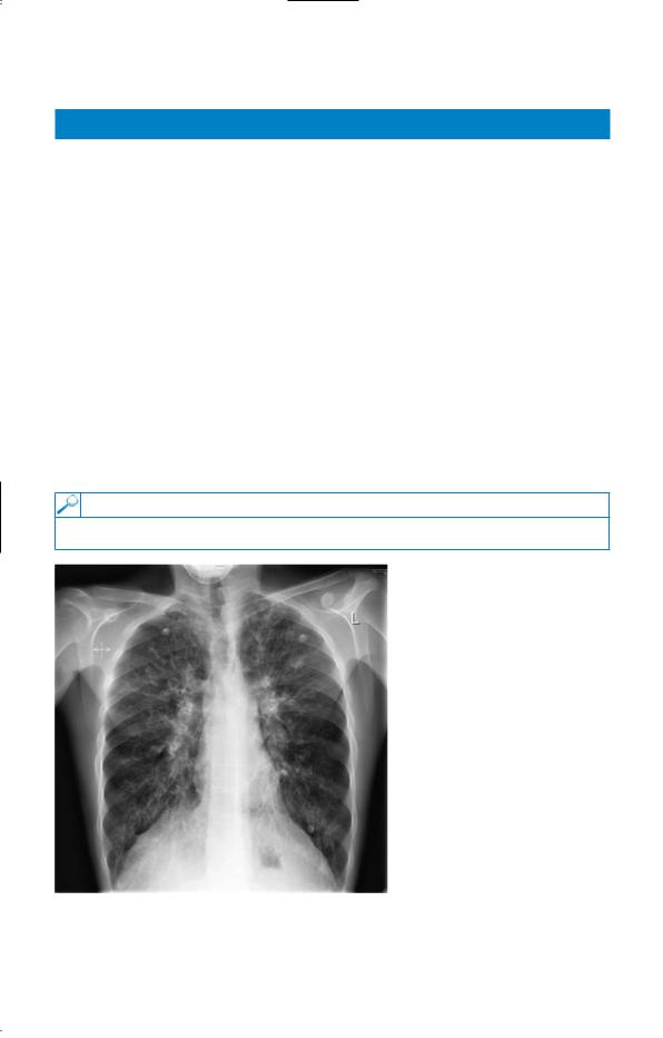
• The chest X-ray is shown in Fig. 3.1.
Figure 3.1 Chest X-ray.
Questions
•What does the X-ray show?
•What is the most likely diagnosis?
•What investigations should be performed?
9

ANSWER 3
The chest X-ray shows abnormal shadowing throughout both lungs, more marked in both upper lobes with some ring shadows and tubular shadows representing thickened bronchial walls. These findings would be compatible with a diagnosis of bronchiectasis. The pulmonary arteries are prominent, suggesting a degree of pulmonary hypertension. The distribution is typical of that found in cystic fibrosis where the changes are most evident in the upper lobes. Most other forms of bronchiectasis are more likely to occur in the lower lobes where drainage by gravity is less effective. High-resolution computed tomography (CT) of the lungs is the best way to diagnose bronchiectasis and to define its extent and distribution. In younger and milder cases of cystic fibrosis, the predominant organisms in the sputum are Haemophilus influenzae and Staphylococcus aureus. Later, as more lung damage occurs, Pseudomonas aeruginosa is a common pathogen. Once present in the lungs in cystic fibrosis, it is difficult or impossible to remove it completely.
Cystic fibrosis should always be considered when there is a story of repeated chest infections in a young person. Although it presents most often below the age of 20 years, diagnosis may be delayed until the 20s, 30s or even 40s in milder cases. Associated problems occur in the pancreas (malabsorption, diabetes), sinuses and liver. It has become evident that some patients are affected more mildly, especially those with the less common genetic variants. These milder cases may only be affected by the chest problems of cystic fibrosis and have little or no malabsorption from the pancreatic insufficiency.
!Differential diagnosis
The differential diagnosis in this young man would be other causes of diffuse bronchiectasis such as agammaglobulinaemia or immotile cilia. Respiratory function should be measured to see the degree of functional impairment. Bronchiectasis in the upper lobes may occur in tuberculosis or in allergic bronchopulmonary aspergillosis associated with asthma.
The common diagnostic test for cystic fibrosis is to measure the electrolytes in the sweat, where there is an abnormally high concentration of sodium and chloride. At the age of 19 years, the sweat test may be less reliable. It is more specific if repeated after the administration of fludrocortisone. An alternative would be to have the potential difference across the nasal epithelium measured at a centre with a special interest in cystic fibrosis. Cystic fibrosis has an autosomal recessive inheritance with the commonest genetic abnormality "F508 found in 85 per cent of cases. The gene is responsible for the protein controlling chloride transport across the cell membrane. The commoner genetic abnormalities can be identified and the current battery of genetic tests identifies well over 95 per cent of cases. However, the absence of "F508 and other common abnormalities would not rule out cystic fibrosis related to the less common genetic variants.
In later stages, lung transplantation can be considered. Since the identification of the genetic abnormality, trials of gene-replacement therapy have begun.
KEY POINTS
•Milder forms of cystic fibrosis may present in adolescence and adulthood.
•Milder forms are often related to less common genetic abnormalities.
•A high-resolution CT scan is the best way to detect bronchiectasis and to define its extent.
10

CASE 4: SHORTNESS OF BREATH
History
A 26-year-old teacher has consulted her general practitioner (GP) for her persistent cough. She wants to have a second course of antibiotics because an initial course of amoxicillin made no difference. The cough has troubled her for 3 months since she moved to a new school. The cough is now disturbing her sleep and making her tired during the day. She teaches games, and the cough is troublesome when going out to the playground and on jogging. In her medical history she had her appendix removed 3 years ago. She had her tonsils removed as a child and was said to have recurrent episodes of bronchitis between the ages of 3 and 6 years. She has never smoked and takes no medication other than an oral contraceptive. Her parents are alive and well and she has two brothers, one of whom has hayfever.
Examination
The respiratory rate is 18/min. Her chest is clear and there are no abnormalities in the nose, pharynx, cardiovascular, respiratory or nervous systems.
INVESTIGATIONS
•Chest X-ray is reported as normal.
•Spirometry is carried out at the surgery and she is asked to record her peak flow rate at home, the best of three readings every morning and every evening for 2 weeks. Spirometry results are as follows:
|
Actual |
Predicted |
FEV1 (L) |
3.9 |
3.6–4.2 |
FVC (L) |
5.0 |
4.5–5.4 |
FER (FEV1/FVC) (%) |
78 |
75–80 |
PEF (L/min) |
470 |
440–540 |
FEV1: forced expiratory volume in 1 s; FVC, forced vital capacity; FER, forced expiratory ratio; PEF, peak expiratory flow.
A peak flow recording is shown in Fig. 4.1.
|
500 |
|
|
|
480 |
|
|
(L/min) |
460 |
|
|
440 |
|
|
|
flow |
|
|
|
420 |
|
|
|
Peak |
|
|
|
400 |
|
|
|
|
|
|
|
|
380 |
|
|
|
360 |
|
|
|
1 |
Days |
10 |
Figure 4.1 Peak flow recording at home over 11 days.
Questions
•What is your interpretation of these findings?
•What do you think is the likely diagnosis and what would be appropriate treatment?
11

ANSWER 4
The peak flow pattern shows a degree of diurnal variation. This does not reach the diagnostic criteria for asthma but it is suspicious. The mean daily variation in peak flow from the recordings is 36 L/min and the mean evening peak flow is 453 L/min, giving a mean diurnal variation of 8 per cent. There is a small diurnal variation in normals and a variation of #15 per cent is diagnostic of asthma. In this patient the label of ‘bronchitis’ as a child was probably asthma. The family history of an atopic condition (hayfever in a brother), and the triggering of the cough by exercise and going out in to the cold also suggest bronchial hyper-responsiveness typical of asthma.
Patients with a chronic persistent cough of unexplained cause should have a chest X-ray. When the X-ray is clear the cough is likely to be produced by one of three main causes in non-smokers. Around half of such cases have asthma or will go on to develop asthma over the next few years. Half of the rest have rhinitis or sinusitis with a post-nasal drip. In around 20 per cent the cough is related to gastro-oesophageal reflux. A small number of cases will be caused by otherwise unsuspected problems such as foreign bodies, bronchial ‘adenoma’, sarcoidosis or fibrosing alveolitis. Cough is a common side-effect in patients treated with angiotensin-converting-enzyme (ACE) inhibitors.
In this patient the diagnosis of asthma was confirmed with an exercise test which was associated with a 25 per cent drop in peak flow after completion of 6 min vigorous exercise. Alternatives would have been another non-specific challenge such as methacholine or histamine, or a therapeutic trial of inhaled steroids.
After the exercise test, an inhaled steroid was given and the cough settled after 1 week. The inhaled steroid was discontinued after 4 weeks and replaced by a $2-agonist to use before exercise. However, the cough recurred with more evident wheeze and shortness of breath, and treatment was changed back to an inhaled steroid with a $2-agonist as needed. If control was not established, the next step would be to check inhaler technique and treatment adherence and to consider adding a long-acting $2-agonist. In some cases, the persistent dry cough associated with asthma may require more vigorous treatment than this. Inhaled steroids for a month or more, or even a 2-week course of oral steroids may be needed to relieve the cough. The successful management of dry cough relies on establishing the correct diagnosis and treating it vigorously.
KEY POINTS
•The three commonest causes of persistent dry cough with a normal chest X-ray are asthma (50 per cent), sinusitis and postnasal drip (25 per cent) and reflux oesophagitis (20 per cent).
•Asthma may present as a cough (cough variant asthma) with little or no airflow obstruction initially, although this develops later.
•Persistent cough with normal chest examination is unlikely to have a bacterial cause or respond to antibiotic treatment.
12
This page intentionally left blank