Материал: 100_Cases_in_Clinical_Medicine
CASE 53: LOSS OF CONSCIOUSNESS
History
An unkempt man of uncertain age, estimated to be 55–65 years, is brought in to hospital by ambulance having been found unconscious on the pavement outside a pub on New Year’s eve. There is no other available history. A used packet of paracetamol and dihydrocodeine is found in one of his pockets but no illicit drugs and no means of identification.
On examination he looks pale and smells of alcohol and urine. There are no signs of head injury and no localizing neurological signs.
Examination
Tendon reflexes are present and equal except the ankle reflexes which are absent. Plantar responses are downgoing. The pupils are equal and reactive and the fundi look normal. The observation chart is completed by the nurse in the emergency department.
INVESTIGATIONS
•Pulse: 82/min
•Blood pressure: 92/56 mmHg
•Temperature: 35.1°C
•Respiratory rate: 12/min
•Oxygen saturation: 95 per cent breathing air
•Glasgow Coma Scale: 10/15
•Urine on catheterization: 450 mL volume; ' sugar; ' blood; no protein
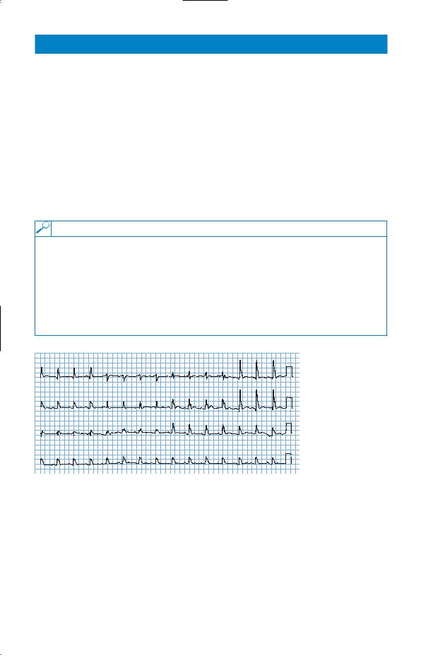
The electrocardiogram (ECG) is shown in Fig. 53.1.
I |
aVR |
V1 |
V4 |
II |
aVL |
V2 |
V5 |
III |
aVF |
V3 |
V6 |
II |
|
|
|
Figure 53.1 Electrocardiogram.
Questions
•What is the likely cause of the problem?
•What investigations and treatment are indicated?
139
ANSWER 53
This man has been unconscious in the open air for an unknown period. Little history is available, but the tablets in his pocket might suggest that he has a problem with a painful condition. There are a number of possible causes for his unconsciousness including a cerebrovascular problem, deliberate or accidental drug overdose, including alcohol poisoning, metabolic or endocrine disturbance or hypothermia.
If this were an overdose, e.g. of dihydrocodeine, the pupils might well be small. The slow respiratory rate could be compatible with an opiate excess suppressing ventilation. The oxygen saturation results show that he is oxygenating himself satisfactorily although it would be sensible to perform blood gases to measure the arterial partial pressure of CO2 (paCO2). It would be appropriate to measure the paracetamol level in the blood and it would be worth giving the opiate antagonist naloxone if there remained a likelihood of overdose. The blood alcohol level should be measured to exclude alcohol poisoning.
Most cerebrovascular problems would be expected to produce some localizing neurological signs on careful examination even in an unconscious patient. There are no such signs here. The absent ankle jerks might be related to his age.
He could have hyperosmolar non-ketotic coma detected by a high glucose and evidence of haemoconcentration. The blood glucose should be measured together with electrolytes and haematology but the single ' of glucose in the urine makes it unlikely that he has hyperglycaemic coma. Liver function and renal function should be measured.
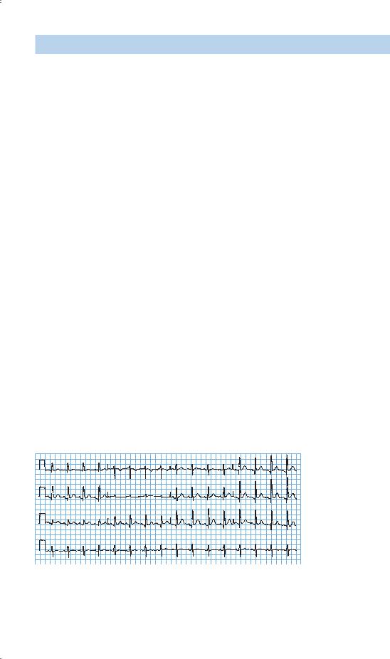
He has a slow respiratory rate, low blood pressure and an ECG which shows a wide QRS complex. The wide complexes on the ECG show an extra deflection at the end of the QRS complex, the J point. This J-wave is characteristic of hypothermia and disappears after rewarming as shown by the subsequent ECG (Fig. 53.2). The pulse rate would often be slower than the 82/min in this man and the ECG may show evidence of a tremor from shivering. The temperature of 35.1°C does not appear excessively low but this may not be reliable if it is not a true core temperature or has been measured with a normal mercury thermometer (mercury thermometers are not reliable at low temperatures). Indeed, in this case, repeat of the rectal temperature measurement with a low-reading thermometer showed a temperature of 30.6°C. No paracetamol was detected in the blood and his alcohol level was low at 11 mg/100 mL.
I |
aVR |
V1 |
V4 |
|
|||
|
|
|
|
II |
aVL |
V2 |
V5 |
III |
aVF |
V3 |
V6 |
|
VI
Figure 53.2 Electrocardiogram of resolved hypothermia.
140

The management of hypothermia is gradual passive rewarming with replacement of fluids by warmed colloids as rewarming takes place. The increase of temperature should be 0.5–1°C per hour. If this is not achieved by covering the patient with blankets, then warmed inspired oxygen, warm intravenous fluids, bladder or peritoneal lavage might be considered. Drugs and physical disturbance should be limited since the myocardium is often irritable and susceptible to arrhythmias.
KEY POINTS
•Hypothyroidism should be considered as a possible contributor to hypothermia.
•Even when alcohol is a cause of unconsciousness, other causes must be excluded.
•The diagnosis of hypothermia requires a thermometer capable of reading low temperatures.
•J-waves on the ECG are specific signs of hypothermia.
•Hypothermia in the elderly is treated by gradual passive rewarming.
141
This page intentionally left blank

CASE 54: TIREDNESS
History
A 22-year-old woman complains of tiredness for 6 months. Her only other symptom is a gradual increase in frequency of bowel movements from once a day in her teens to two to three times daily. She has no abdominal pain and has no change in appetite. She says that the bowel movements can be difficult to flush away on occasions but this is not a consistent problem. She is a non-smoker and drinks rarely. She has been a vegetarian for 5 years but eats dairy foods and fish regularly. She thinks that her grandmother, who lived in Ireland, had some bowel problems but she died 3 years ago, aged 68. She is an infantschool teacher and spends a lot of her spare time in keep-fit classes and routines at a local gym. She enjoys her work and socializes regularly with a wide circle of friends.
Examination
She is 1.62 m (5 ft 4 in) tall and weighs 49 kg. She looks a little pale and thin. Examination of her abdomen showed no abnormalities and there are no other significant abnormalities to find in any other system.
INVESTIGATIONS
|
|
Normal |
Haemoglobin |
10.7 g/dL |
11.7–15.7 g/dL |
Mean corpuscular volume (MCV) |
98 fL |
80–99 fL |
White cell count |
6.5 % 109/L |
3.5–11.0 % 109/L |
Platelets |
247 % 109/L |
150–440 % 109/L |
Red cell folate |
44 mg/L |
#160 mg/L |
Vitamin B12 |
280 ng/L |
176–925 ng/L |
Thyroid-stimulating hormone |
3.5 mU/L |
0.3–6.0 mU/L |
Free thyroxine |
12.9 pmol/L |
9.0–22.0 pmol/L |
The blood film is reported as a dimorphic film with remnants of nuclear material (Howell–Jolly bodies) in some of the red blood cells.
Questions
•How do you interpret these findings?
•What is the likely diagnosis and how might this be confirmed?
143