Материал: 100_Cases_in_Clinical_Medicine

ANSWER 54
The most likely diagnosis is malabsorption from coeliac disease. The report of a dimorphic blood film means that there are both small and large cells. This suggests that the anaemia is caused by a combination of the folate deficiency indicated by the red cell folate and by iron deficiency. The Howell–Jolly bodies are dark blue regular inclusions in the red cells which are typically found in the blood of patients after splenectomy, or are associated with the splenic atrophy which is characteristic of coeliac disease. In coeliac disease, there is a sensitivity to dietary gluten, a water-insoluble protein found in many cereals. The proximal small bowel is the main site involved with loss of villi and an inflammatory infiltrate causing reduced absorption.
The MCV is at the upper limit of normal.
!Causes of macrocytosis in the blood film
•Folate deficiency
•Vitamin B12 deficiency
•Excessive alcohol consumption
•Hypothyroidism
•Certain drugs, e.g. azathioprine, methotrexate
•Primary acquired sideroblastic anaemia and myelodysplastic syndromes
Coeliac disease is made more likely by a possible positive family history and the origin from Ireland where coeliac disease is four times as common as in the rest of the UK. Other diagnoses which might be considered are anorexia nervosa (her age and sex, commitment to exercise); she does not appear depressed (a common cause of weight loss and bowel disturbance) and the laboratory findings clearly indicate physical disease.
Diagnosis of coeliac disease can be confirmed by endoscopy at which a biopsy can be taken from the distal duodenum. Typically this will show complete villus atrophy. Antigliadin antibodies are usually positive and can be a useful screening test. The treatment is a glutenfree diet with a repeat of the biopsy some months later to show improvement in the height of the villi in the small bowel. In some cases, temporary treatment with steroids may be needed to help recovery. Another common cause of failure to recover the villus architecture is poor compliance to the difficult dietary constraints.
KEY POINTS
•Howell–Jolly bodies are characteristic of hyposplenism.
•Coeliac disease can present at any age with non-specific symptoms; absence of abdominal pain and/or steatorrhoea are not unusual.
•Typical features of fat malabsorption may not be evident if the patient eats a diet with little or no fat intake.
144
CASE 55: RECURRENT CHEST INFECTIONS
History
A 45-year-old woman is admitted to hospital with pneumonia. She has had three episodes of cough, fever and purulent sputum over the last 6 months. One of these was associated with right-sided pleuritic chest pain. These have been treated at home by her general practitioner (GP). In addition she has a 5-year history of difficulty with swallowing. Initially this was mild but it has become progressively worse. She says that food seems to stick in the low retrosternal area. This applies to all types of solid food. She has lost 5 kg in weight over the last 2 months. Sometimes the difficulty with swallowing seems to improve during a meal. Recently she has had trouble with regurgitation and vomiting of recognizable food.
Three years ago her GP arranged for an outpatient upper gastrointestinal endoscopy which was normal. She was reassured, but the problem has increased in severity. There is no other relevant medical history or family history. She lived in the north-west coast of the United States for 4 years up until 10 years ago. She works as a shop assistant. She has never smoked and drinks less than 5 units of alcohol each week. There has been no disturbance of micturition. She has always tended to be constipated and this has been a little worse recently.
Examination
She looks thin. In the respiratory system there are some crackles at the right base. There are no abnormalities to find in the cardiovascular system, abdomen or other systems.
INVESTIGATIONS
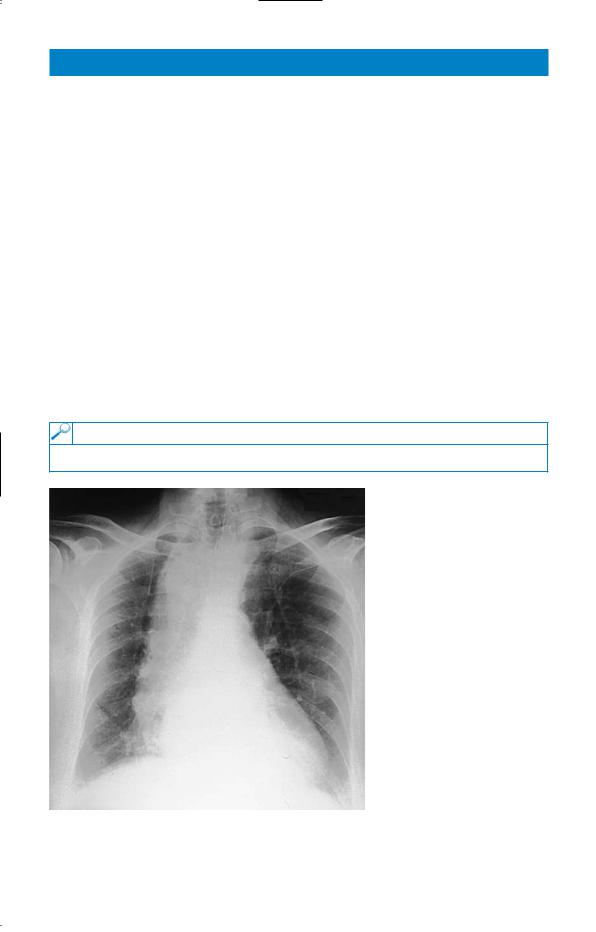
Her chest X-ray is shown in Fig. 55.1.
Figure 55.1 Chest X-ray. (reproduced with the kind permission from Curtis and Whitehouse,
Radiology for the MRCP,
Arnold, 1998.)
Questions
•What is the likely diagnosis?
•How would you establish this?
145

ANSWER 55
The likely diagnosis is achalasia of the cardia, a primary neurological disturbance of the nerve plexuses at the lower end of the oesophagus. The X-ray shows a dilated fluid-filled oesophagus with no visible gastric air bubble. Endoscopy may be normal in the early stages as in this case. The oesophagus has now dilated and there has been spill-over of stagnant food into the lungs giving her the episodes of repeated respiratory infections. Such aspiration is most likely to affect the right lower lobe because of the more vertical right main bronchus, although the result of aspiration at night may depend on the position of the patient. The dysphagia is often variable early on. It tends to be present for all foods, indicating a motility problem, and there may initially be some relief from the mechanical load as the oesophagus fills. Dysphagia for bulky, solid foods first usually indicates an obstructive lesion.
The diagnosis can be made at this stage by a barium swallow showing the dilated oesophagus. Earlier it may require careful cine-radiology with a bolus of food impregnated with barium, or oesophageal motility studies using a catheter fitted with a number of pressure sensors to detect the abnormal motility of the oesophageal muscle.
A similar condition can be produced by the protozoan parasite Trypanosoma cruzi (Chagas’ disease), but this is limited to South and Central America and would not be relevant to her stay in the north-west United States.
Other common causes of dysphagia are benign oesophageal structures from acid reflux, malignant structures, external compression or an oesophageal pouch. Achalasia may be managed by muscle relaxants when mild, but often requires treatment to disrupt the lower oesophageal muscle by dilatation or surgery.
KEY POINTS
•The subjective site of blockage in dysphagia may not reflect accurately the level of the obstruction.
•Persistent dysphagia without explanation needs investigation by barium swallow or endoscopy.
146

CASE 56: GENERALIZED ITCHING
History
A 63-year-old man presents to his general practitioner (GP) complaining that over the past 3 months he has developed severe generalized itching. This is particularly severe after a hot bath. He has also noticed frequent headaches, dizziness and tinnitus. He has been hypertensive for 10 years. He smokes 35 cigarettes a day and drinks about 25 units of alcohol per week. He takes bendroflumethiazide for his hypertension. He works as a chef, is divorced and lives alone.
Examination
His face looks plethoric. He has no rash. There are no signs of chronic liver disease and no lymphadenopathy. His pulse is 84/min regular and blood pressure 162/104 mmHg. Examination of his heart and respiratory system is normal. In his abdomen the only abnormality is that his spleen is palpable 4 cm below the left costal margin. Neurological examination is normal.
INVESTIGATIONS
|
|
Normal |
Haemoglobin |
18.6 g/dL |
13.3–17.7 g/dL |
Mean corpuscular volume (MCV) |
88 fL |
80–99 fL |
White cell count |
14.2 % 109/L |
3.9–10.6 % 109/L |
Platelets |
598 % 109/L |
150–440 % 109/L |
Sodium |
137 mmol/L |
135–145 mmol/L |
Potassium |
4.6 mmol/L |
3.5–5.0 mmol/L |
Urea |
6.4 mmol/L |
2.5–6.7 mmol/L |
Creatinine |
84 &mol/L |
70–120 &mol/L |
Bilirubin |
12 mmol/L |
3–17 mmol/L |
Alanine transaminase |
24 IU/L |
5–35 IU/L |
Alkaline phosphatase |
68 IU/L |
30–300 IU/L |
Urinalysis: no protein; no blood |
|
|
Questions
•What is the likely diagnosis?
•What are the major causes of generalized itching without a rash?
•How would you investigate and manage this patient?
147

ANSWER 56
This patient has polycythaemia rubra vera. This is due to abnormal proliferation of red cell precursors derived from a single haematopoietic progenitor cell with the capacity for differentiation down red cell, white cell and platelet lines. As a result, there is an increase in haemoglobin, white cell count and platelet level. Patients may present with a thrombotic event or with symptoms due to increased blood viscosity such as headaches, tinnitus and blurred vision. There is an increased bleeding tendency. Splenomegaly is common. Severe pruritus is characteristic and is particularly related to warmth occurring on getting into a warm bed or bath.
!Conditions associated with generalized pruritus without a rash
•Obstructive jaundice due to bile salt retention
•Iron deficiency
•Lymphoma
•Carcinoma, especially bronchial
•Chronic renal failure, partially due to phosphate retention
This patient should be referred to a haematology unit for investigation. It is important to exclude relative polycythaemia due to dehydration from diuretic and alcohol use. The red cell mass will be raised in polcythaemia rubra vera, but normal in relative polycythaemia. The following causes of secondary polycythaemia must be excluded:
•chronic lung disease with hypoxia
•cyanotic congenital heart disease
•renal cysts, tumours, renal transplants
•hepatoma, cerebellar haemangioblastoma, uterine fibroids
•Cushing’s disease.
The erythropoietin level is low in polycythaemia rubra vera and high in secondary polycythaemia. Pulse oximetry or arterial blood gases should be performed to exclude hypoxia. The leucocyte alkaline phosphatase level is also raised in polycythaemia rubra vera.
The patient should be venesected until the haematocrit is within the normal range. A variety of agents can be used to keep the haematocrit down: 32P, hydroxyurea and busulphan. The disease may transform into acute leukaemia or myelosclerosis.
KEY POINTS
• Severe generalized pruritus is a characteristic symptom of polycythaemia rubra vera.
148