Материал: 100_Cases_in_Clinical_Medicine
This page intentionally left blank

CASE 57: COUGH AND JOINT PAINS
History
A 29-year-old man presents with a cough and some mild aches in the hands, wrists and ankles. The symptoms have been present for 2 months and have increased slightly over that time. Six weeks before he had some soreness of his eyes, which resolved in 1 week.
The cough has been non-productive. He had noticed some skin lesions on the edge of the hairline and around his nostrils. Previously he had been well apart from an appendicectomy at the age of 17 years.
He was born in Trinidad and came to the UK at the age of 4 years. His two brothers and parents are well. He does not smoke, is teetotal and takes no recreational drugs. He works as a messenger and took regular exercise until the last few weeks.
Examination
There is no deformity of the joints and no evidence of any acute inflammation. In the respiratory and cardiovascular system there are no abnormal findings. In the skin there are some slightly raised areas on the edge of the hairline posteriorly and at the ala nasae. They are a little lighter than the rest of the skin.
INVESTIGATIONS
|
|
Normal |
Haemoglobin |
13.5 g/dL |
13.0–17.0 g/dL |
Mean corpuscular volume (MCV) |
88 fL |
80–99 fL |
White cell count |
8.5 % 109/L |
3.5–11.0 % 109/L |
Platelets |
264 % 109/L |
150–440 % 109/L |
Erythrocyte sedimentation rate (ESR) |
34 mm |
!10 mm/h |
Sodium |
140 mmol/L |
135–145 mmol/L |
Potassium |
4.0 mmol/L |
3.5–5.0 mmol/L |
Urea |
3.6 mmol/L |
2.5–6.7 mmol/L |
Creatinine |
74 &mol/L |
70–120 &mol/L |
Bilirubin |
14 mmol/L |
3–17 mmol/L |
Alkaline phosphatase |
84 IU/L |
30–300 IU/L |
Alanine aminotransferase |
44 IU/L |
5–35 IU/L |
Calcium |
2.69 mmol/L |
2.12–2.65 mmol/L |
Phosphate |
1.20 mmol/L |
0.8–1.45 mmol/L |
The chest X-ray is shown in Fig. 57.1. |
|
|
150
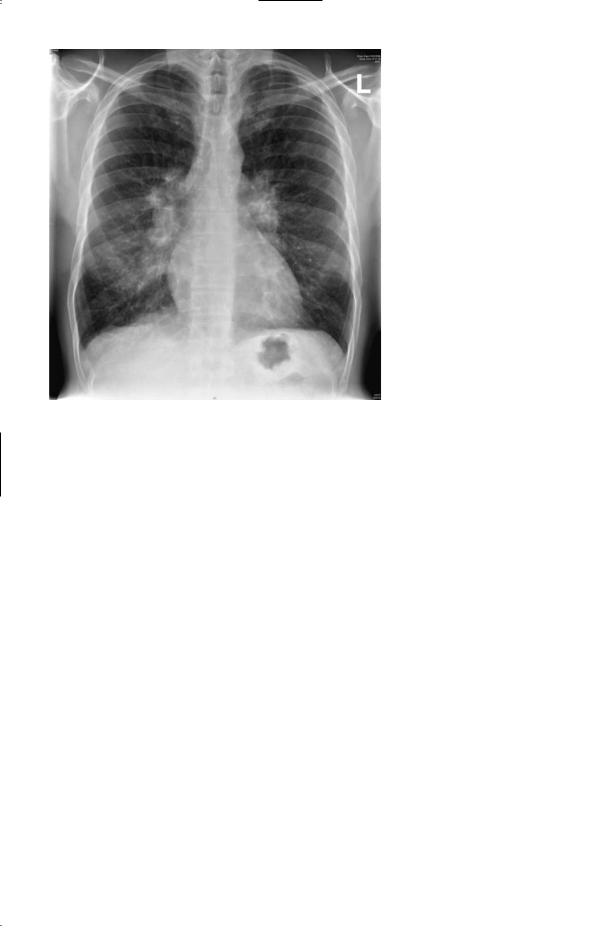
Figure 57.1 Chest X-ray.
Questions
•What is the likely diagnosis?
•How might this be confirmed?
151

ANSWER 57
The likely diagnosis is sarcoidosis. The age is typical and sarcoidosis is more common in those of African-Caribbean origin. The chest X-ray shows bilateral hilar lymphadenopathy. The blood results show a slightly raised calcium level which is related to vitamin D sensitivity in sarcoidosis where the granulomas hydroxylate 25-hydroxycholecalciferol to 1,25dihydroxycholecalciferol. The ESR is raised and some of the liver enzymes are around the upper limit of normal. The skin lesions at the hairline and the nostrils are typical sites for sarcoid skin problems. The eye trouble 6 weeks earlier might also have been a manifestation of sarcoidosis, which can cause both anterior and posterior uveitis.
An alternative diagnosis which might explain the findings is tuberculosis. Tuberculosis can also cause hypercalcaemia although this is much less common than in sarcoid. Tumours, especially lymphoma, might give this X-ray appearance but would not explain the other findings. The arthralgia (pains with no evidence of acute inflammation or deformity on examination) can occur in sarcoid or tuberculosis but again they are commoner in sarcoid. The ESR is non-specific. Arthralgia without deformity in an African-Caribbean man raises the possibility of systemic lupus erythematosus (SLE), but this would be much commoner in women and would not cause bilateral hilar lymphadenopathy.
He is likely to have had BCG (bacille Calmette–Guèrin) vaccination at school at around the age of 12 years, giving a degree of protection against tuberculosis. A tuberculin test should be positive after BCG, strongly positive in most cases of tuberculosis and negative in 80 per cent of cases of sarcoidosis. The serum level of angiotensin-converting enzyme would be raised in over 80 per cent of cases of sarcoidosis but often in tuberculosis also; the granuloma cells secrete this enzyme. A computed tomography (CT) scan of the chest will confirm the extent of the lymphadenopathy and show whether there is any involvement of the lung parenchyma. Histology of affected tissue would confirm the clinical diagnosis. This might be obtained by a skin biopsy of one of the lesions. A bronchial or transbronchial lung biopsy at fibreoptic bronchoscopy would be another means of obtaining diagnostic histology. In patients with a cough and sarcoidosis the bronchial mucosa itself often looks abnormal, and biopsy will provide the diagnosis. Lung function tests and electrocardiogram (ECG) should be performed as a baseline if the diagnosis is confirmed.
Steroid treatment would not be necessary for the hilar lymphadenopathy alone, but would be indicated for the hypercalcaemia and possibly for the systemic symptoms.
KEY POINTS
•Sarcoidosis is commoner in African-Caribbeans.
•Typical sites for skin lesions are around the nose and the hairline.
•Sarcoidosis is a systemic disease and can affect most parts of the body.
152

CASE 58: THIRST AND FREQUENCY
History
A 63-year-old woman is referred to a nephrologist for investigation of polyuria. About 4 weeks ago she developed abrupt-onset extreme thirst and polyuria. She is getting up to pass urine five times a night. Over the past 3 months she has felt generally unwell and noted pain in her back. She has lost 3 kg in weight over this time. She also has a persistent frontal headache associated with early morning nausea. The headache is worsened by coughing or lying down. Eight years previously she had a left mastectomy and radiotherapy for carcinoma of the breast. She is a retired civil servant who is a non-smoker and drinks 10 units of alcohol per week. She is on no medication.
Examination
She is thin and her muscles are wasted. Her pulse rate is 72/min, blood pressure 120/84 mmHg, jugular venous pressure is not raised, heart sounds are normal and she has no peripheral oedema. Examination of her respiratory, abdominal and neurological systems is normal. Her fundi show papilloedema.
INVESTIGATIONS
|
|
Normal |
Haemoglobin |
12.2 g/dL |
11.7–15.7 g/dL |
Mean corpuscular volume (MCV) |
85 fL |
80–99 fL |
White cell count |
6.7 % 109/L |
3.5–11.0 % 109/L |
Platelets |
312 % 109/L |
150–440 % 109/L |
Sodium |
142 mmol/L |
135–145 mmol/L |
Potassium |
3.8 mmol/L |
3.5–5.0 mmol/L |
Bicarbonate |
26 mmol/L |
24–30 mmol/L |
Urea |
4.2 mmol/L |
2.5–6.7 mmol/L |
Creatinine |
68 &mol/L |
70–120 &mol/L |
Glucose |
4.2 mmol/L |
4.0–6.0 mmol/L |
Albumin |
38 g/L |
35–50 g/L |
Calcium |
2.75 mmol/L |
2.12–2.65 mmol/L |
Phosphate |
1.2 mmol/L |
0.8–1.45 mmol/L |
Bilirubin |
12 mmol/L |
3–17 mmol/L |
Alanine transaminase |
35 IU/L |
5–35 IU/L |
Alkaline phosphatase |
690 IU/L |
30–300 IU/L |
Urinalysis: no protein; no blood |
|
|
Questions
•What is the likely cause of her polyuria?
•How would you investigate and manage this patient?
153