Материал: 100_Cases_in_Clinical_Medicine

ABDOMEN
CASE 5: ACUTE ABDOMINAL PAIN
History
A 56-year-old woman presents to the emergency department complaining of abdominal pain. Twenty-four hours previously she developed a continuous pain in the upper abdomen which has become progressively more severe. The pain radiates into the back. She feels nauseated and alternately hot and cold. Her past medical history is notable for a duodenal ulcer which was successfully treated with Helicobacter eradication therapy 5 years earlier. She smokes 15 cigarettes a day, and shares a bottle of wine each evening with her husband.
Examination
The patient looks unwell and dehydrated. She weighs 115 kg. She is febrile, 38.5°C, her pulse is 108/min and blood pressure 124/76 mmHg. Cardiovascular and respiratory system examination is normal. She is tender in the right upper quadrant and epigastrium, with guarding and rebound tenderness. Bowel sounds are sparse.
INVESTIGATIONS
|
|
Normal |
Haemoglobin |
14.7 g/dL |
11.7–15.7 g/dL |
White cell count |
19.8 % 109/L |
3.5–11.0 % 109/L |
Platelets |
239 % 109/L |
150–440 % 109/L |
Sodium |
137 mmol/L |
135–145 mmol/L |
Potassium |
4.8 mmol/L |
3.5–5.0 mmol/L |
Urea |
8.6 mmol/L |
2.5–6.7 mmol/L |
Creatinine |
116 &mol/L |
70–120 &mol/L |
Bilirubin |
19 &mol/L |
3–17 &mol/L |
Alkaline phosphatase |
58 IU/L |
30–300 IU/L |
Alanine aminotransferase (AAT) |
67 IU/L |
5–35 IU/L |
Gamma-glutamyl transpeptidase |
72 IU/L |
11–51 IU/L |
C-reactive protein (CRP) |
256 mg/L |
!5 mg/L |
A plain abdominal X-ray is shown in Fig. 5.1.
14
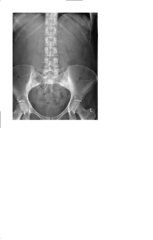
Figure 5.1 Plain abdominal
X-ray.
Questions
•What is the most likely diagnosis?
•How would you manage this patient?
15

ANSWER 5
This woman has acute cholecystitis. Cholecystitis is most common in obese, middle-aged women, and classically is triggered by eating a fatty meal. Cholecystitis is usually caused by a gallstone impacting in the cystic duct. Continued secretion by the gallbladder leads to increased pressure and inflammation of the gallbladder wall. Bacterial infection is usually by Gram-negative organisms and anaerobes. Ischaemia in the distended gallbladder can lead to perforation causing either generalized peritonitis or formation of a localized abscess. Alternatively the stone can spontaneously disimpact and the symptoms spontaneously improve. Gallstones can get stuck in the common bile duct leading to cholangitis or pancreatitis. Rarely, gallstones can perforate through the inflamed gallbladder wall into the small intestine and cause intestinal obstruction (gallstone ileus). The typical symptom is of sudden-onset right upper quadrant abdominal pain which radiates into the back. In uncomplicated cases the pain improves within 24 h. Fever suggests a bacterial infection. Jaundice usually occurs if there is a stone in the common bile duct. There is usually guarding and rebound tenderness in the right upper quadrant (Murphy’s sign).
In this patient the leucocytosis and raised CRP are consistent with acute cholecystitis. If the serum bilirubin and liver enzymes are very deranged, acute cholangitis due to a stone in the common bile duct should be suspected. The abdominal X-ray is normal; the majority of gallstones are radiolucent and do not show on plain films.
!Differential diagnosis
The major differential diagnoses of acute cholecystitis include perforated peptic ulcer, acute pancreatitis, acute hepatitis, subphrenic abscess, retrocaecal appendicitis and perforated carcinoma or diverticulum of the hepatic flexure of the colon. Myocardial infarction or right lower lobe pneumonia may also mimic cholecystitis.
This patient should be admitted under the surgical team. Serum amylase should be measured to rule out pancreatitis. Blood cultures should be taken. Chest X-ray should be performed to exclude pneumonia, and erect abdominal X-ray to rule out air under the diaphragm which occurs with a perforated peptic ulcer. An abdominal ultrasound will show inflammation of the gallbladder wall. The patient should be kept nil by mouth, given intravenous fluids and commenced on intravenous cephalosporins and metronidazole. The patient should be examined regularly for signs of generalized peritonitis or cholangitis. If the symptoms settle down the patient is normally discharged to be readmitted in a few weeks once the inflammation has settled down to have a cholecystectomy.
KEY POINTS
•Acute cholecystitis typically causes right upper quadrant pain and a positive Murphy’s sign.
•Potential complications include septicaemia and peritonitis.
16

CASE 6: WEIGHT LOSS
History
A 66-year-old woman, a retired nurse, consults her general practitioner (GP) with a 4-month history of tiredness, slight breathlessness on exertion and loss of weight from 71 to 65 kg. Her appetite is unchanged and normal, she has no nausea or vomiting, but over the last 2 months she has had an altered bowel habit with constipation alternating with her usual and normal pattern. She has not seen any blood in her faeces and has had no abdominal pain. There is no relevant past or family history, and she is on no medication.
She has smoked 20 cigarettes daily for 48 years and drinks 20–28 units of alcohol a week.
Examination
She has slight pallor but otherwise looks well. No lymphadenopathy is detected, and her breasts, thyroid, heart, chest and abdomen, including rectal examination, are all normal. The blood pressure is 148/90 mmHg.
INVESTIGATIONS
|
|
Normal |
Haemoglobin |
10.1 g/dL |
11.7–15.7 g/dL |
Mean corpuscular volume (MCV) |
76 fL |
80–99 fL |
White cell count |
4.9 % 109/L |
3.5–11.0 % 109/L |
Platelets |
277 % 109/L |
150–440 % 109/L |
Sodium |
142 mmol/L |
135–145 mmol/L |
Potassium |
4.4 mmol/L |
3.5–5.0 mmol/L |
Urea |
5.2 mmol/L |
2.5–6.7 mmol/L |
Creatinine |
106 &mol/L |
70–120 &mol/L |
Urinalysis: no protein, no blood |
|
|
Blood film shows a microcytic hypochromic picture.
Questions
•What is the likeliest diagnosis?
•How would you investigate the patient?
17

ANSWER 6
The microcytic, hypochromic anaemia and the altered bowel habit, the only symptom referable to the gastrointestinal tract, point to a carcinoma of the colon, which would also explain her weight loss. A barium enema revealed a neoplasm in the sigmoid colon, confirmed by colonoscopy and biopsy. Chest X-ray and abdominal ultrasound showed no pulmonary metastases and no intra-abdominal lymphadenopathy or hepatic metastases respectively.
She proceeded to a sigmoid colectomy and end-to-end anastamosis, and was regularly followed-up for any evidence of recurrence. Histology showed a grade I tumour.
Carcinoma of the colon is increasing in frequency. If it presents at an early stage then the prospect for cure is good. Rectal bleeding, alteration in bowel habit for longer than 1 month at any age, or iron-deficient anaemia in men or postmenopausal women are indications for investigation of the gastrointestinal tract.
Smoking is a risk factor for carcinoma of the colon.
KEY POINTS
•Carcinoma of the colon can present with few or no symptoms or signs in the gastrointestinal tract.
•Carcinoma of the colon must be considered as a cause of iron-deficient anaemia.
18