Материал: 100_Cases_in_Clinical_Medicine
CASE 28: CHRONIC CHEST PAIN
History
A 25-year-old woman is complaining of chest pain. This had been present for 2 years on and off. The pain settled for a period of 6 months but it has returned over the last 10 months. The pain is usually on the left side of the chest, radiating to the left axilla. She describes it as a tight or gripping pain which lasts for anything from 5 to 30 min at a time. It can come on at any time, and is often related to exercise but it has occurred at rest on some occasions, particularly in the evenings. The pain is usually associated with shortness of breath. It makes her stop whatever she is doing and she often feels faint or dizzy with the pain. Occasionally palpitations come on after the start of the pain. Detailed questioning about the palpitations indicates that they are a sensation of a strong but steady heart beat.
In her previous medical history she had her appendix removed at the age of 15 years. At the age of 30 years she was investigated for an irregular bowel habit and abdominal pain but no specific diagnosis was arrived at. These symptoms still trouble her. She has seasonal rhinitis. Two years ago she visited a chemist and had her cholesterol level measured; the result was 4.1 mmol/L. In her family history her grandfather died of a myocardial infarction, a year previously, aged 77 years. Several members of her family have hay fever or asthma. She works as a medical secretary. She is married and has no children.
Examination
On examination, she has a blood pressure of 102/65 mmHg and pulse of 78/min which is regular. The heart sounds are normal. There is some tenderness on the left side of the chest, to the left of the sternum and in the left submammary area. The respiratory rate is 22/min. No abnormalities were found on examination of the lungs. She is tender in the left iliac fossa.
INVESTIGATIONS
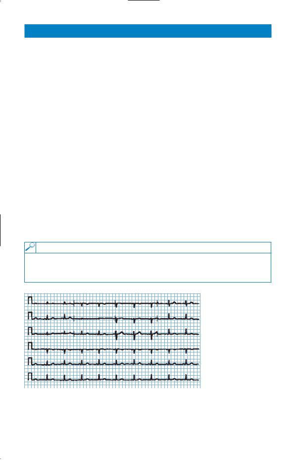
•Her electrocardiogram (ECG) is shown in Fig. 28.1.
•She asked to be referred for a coronary arteriogram to rule out significant coronary artery disease.
I |
aVR |
V1 |
V4 |
|
|||
II |
aVR |
V2 |
V5 |
|
|||
III |
aVF |
V3 |
V6 |
|
|||
V1 |
|
|
|
II |
|
|
|
V5 |
|
|
Figure 28.1 |
|
|
Electrocardiogram. |
|
|
|
|
Question
• What should be done?
79

ANSWER 28
The ECG shown is normal. The pain does not have the characteristics of ischaemic heart disease. On the basis of the information given here it would be reasonable to explore her anxieties and to reassure the patient that this is very unlikely to represent coronary artery disease and to assess subsequently the effects of that reassurance. It may well be that she is anxious about the death of her grandfather from ischaemic heart disease. He may have had symptoms before his death which were related to her anxieties. From a risk point of view her grandfather’s death at the age of 77 with no other affected relatives is not a relevant risk factor. She has expressed anxiety already by having the cholesterol measured (and found to be normal).
She has a history which is suspicious of irritable bowel syndrome with persistent pain, irregular bowel habit and normal investigations. Ischaemic chest pain is usually central and generally reproducible with the same stimuli. The associated shortness of breath may reflect overventilation coming on with the pain and giving her dizziness and palpitations.
The characteristics of the pain and associated shortness of breath should be explored further. Asthma can sometimes be described as tightness or pain in the chest, and she has seasonal rhinitis and a family history of asthma. Gastrointestinal causes of pain such as reflux oesophagitis are unlikely in view of the site and relationship on occasions to exercise. The length of the history excludes other causes of acute chest pain such as pericarditis.
The problem of embarking on tests is that there is no simple screening test which can definitively rule out significant coronary artery disease. Too many investigations may reinforce her belief in her illness and false-positive findings do occur and may exacerbate her anxieties. However, if the patient could not be simply reassured it might be appropriate to proceed with an exercise stress test or a thallium scan to look for areas of reversible ischaemia on exercise or other stress. A coronary arteriogram would not be appropriate without other information to indicate a higher degree of risk of coronary artery disease.
KEY POINTS
•Ischaemic heart disease characteristically causes central rather than left-sided chest pain.
•The resting ECG may show signs of ischaemia or previous infarction but is not a very sensitive test for ischaemic heart disease.
80

CASE 29: OVERDOSE?
History
A 30-year-old woman is brought up to the emergency department at 2 pm by her husband. He is worried that she has taken some tablets in an attempt to harm herself. She has a history suggestive of depression since the birth of her son 3 months earlier. She has been having some counselling since that time but has not been on any medication. The previous evening about 10 pm she told her husband that she was going to take some pills and locked herself in the bathroom. Two hours later he persuaded her to come out and she said that she had not taken anything. They went to bed but he has brought her up now because she has complained of a little nausea and he is worried that she might have taken something when she was in the bathroom. The only tablets in the house were aspirin, paracetamol and temazepam which he takes occasionally for insomnia.
She complains of a little nausea although she has not vomited. She has had a little abdominal discomfort. There is no relevant previous medical or family history of note. She worked as a social worker until 30 weeks of the pregnancy.
Examination
On examination she is mentally alert. She says that she feels sad. Her pulse is 76/min, blood pressure is 124/78 mmHg and respiratory rate is 16/min. There is some mild abdominal tenderness in the upper abdomen but nothing else abnormal to find.
INVESTIGATIONS
|
|
Normal |
Haemoglobin |
12.7 g/dL |
11.7–15.7 g/dL |
Mean corpuscular volume (MCV) |
87 fL |
80–99 fL |
White cell count |
6.8 % 109/L |
3.5–11.0 % 109/L |
Platelets |
230 % 109/L |
150–440 % 109/L |
Prothrombin time |
18 s |
10–14 s |
Sodium |
139 mmol/L |
135–145 mmol/L |
Potassium |
3.8 mmol/L |
3.5–5.0 mmol/L |
Urea |
4.6 mmol/L |
2.5–6.7 mmol/L |
Creatinine |
81 &mol/L |
70–120 &mol/L |
Alkaline phosphatase |
88 IU/L |
30–300 IU/L |
Alanine aminotransferase (AAT) |
37 IU/L |
5–35 IU/L |
Gamma-glutamyl transpeptidase |
32 IU/L |
11–51 IU/L |
Glucose |
5.1 mmol/L |
4.0–6.0 mmol/L |
Question
• What should the management be now?
81

ANSWER 29
It is not evident from the history that the patient herself has been asked about any tablets or other agents she has taken. This would be an important area to be sure of. Of the three agents mentioned, the only one likely to be relevant is paracetamol. Aspirin and temazepam would be likely to produce more symptoms in less than 14 h if they have been taken in significant quantity. However, the salicylate level should certainly be measured; in this case it was not raised. In the absence of drowsiness at this time, it is not necessary to consider temazepam any further.
Paracetamol overdose causes hepatic and renal damage, and can lead to death from acute liver failure. The severity of paracetamol poisoning is dose related with a dose of 15 g being serious in most patients. Patients with pre-existing liver disease and those with a high alcohol intake may be susceptible to smaller overdoses.
The only significant abnormality on the blood tests is a slightly high prothrombin time and minimally raised alanine aminotransferase (AAT). The prothrombin time increase (expressed alternatively as the international normalized ratio or INR) is a signal that a paracetamol overdose is likely. It is often the first test to become abnormal when there is liver damage from paracetamol overdose. If the INR is abnormal at 24 h, then a significant problem is very likely. There are few symptoms in the first 24 h except perhaps nausea, vomiting and abdominal discomfort. This may be associated with tenderness over the liver. The liver function tests usually become abnormal after the first 24 h. Maximum liver damage, as assessed by raised liver enzymes and INR occurs at days 3–4 after overdose. Acute liver failure may develop between days 3 and 5, and renal failure occurs in about 25 per cent of patients with severe hepatic damage. Rarely, renal failure can occur without serious liver damage.
The paracetamol level should be measured urgently; it was found to be high. The evidence of early liver damage from the INR would in itself suggest that treatment with acetylcysteine would be appropriate. The earlier this is used the better but it is certainly still worthwhile 16 h after the ingestion. In this case a level of paracetamol of 64 mg/L confirmed that treatment was appropriate and that the risk of severe liver damage was high. Further advice can always be obtained by ringing one of the national poisons information services. The electrolyte, renal and liver function tests and the clotting studies should be monitored carefully over the first few days, and referral to a liver unit considered if there is marked liver dysfunction. Patients with fulminant hepatic failure are considered for urgent liver transplantation.
The other areas that need to be addressed in this case are the mental state and the safety and care of the son and any other children. This is a serious drug overdose. She should be seen by a psychiatrist or other appropriately trained health worker. The question of any possible risk to the baby should be evaluated before she returns home.
KEY POINTS
•Intravenous acetylcysteine and oral methionine are effective treatments for paracetamol overdose if started early enough.
•Paracetamol levels can be used to predict problems and guide treatment if the time since overdose is known.
•Paracetamol overdose should be suspected in any patient admitted with deranged liver function tests and clotting, if no obvious alternative cause is apparent.
82

CASE 30: AMENORRHOEA
History
A 23-year-old actress presents to her general practitioner (GP) complaining that she has not had a menstrual period for 5 months. She started having periods aged 13 years and previously they had been regular. On direct questioning she states that she has lost 8 kg in weight over the past year although she says her appetite is good. She has had no serious medical illnesses. Currently she is out of work. She split up from her boyfriend 1 year ago. She drinks 10 units of alcohol per week and is a non-smoker. She is taking no regular medication.
Examination
She has lost muscle mass especially on her limbs and buttocks. She is 1.7 m (5 ft 9 in) tall and weighs only 41 kg. She has excessive hair growth over her cheeks, neck and forearms. Her pulse rate is 52/min regular, blood pressure 96/60 mmHg. Examination is otherwise normal.
INVESTIGATIONS
|
|
Normal |
Haemoglobin |
15.2 g/dL |
11.7–15.7 g/dL |
Mean corpuscular volume (MCV) |
84 fL |
80–99 fL |
White cell count |
4.1 % 109/L |
3.5–11.0 % 109/L |
Platelets |
365 % 109/L |
150–440 % 109/L |
Sodium |
136 mmol/L |
135–145 mmol/L |
Potassium |
2.9 mmol/L |
3.5–5.0 mmol/L |
Chloride |
90 mmol/L |
95–105 mmol/L |
Bicarbonate |
33 mmol/L |
24–30 mmol/L |
Urea |
4.2 mmol/L |
2.5–6.7 mmol/L |
Creatinine |
43 &mol/L |
70–120 &mol/L |
Glucose |
5.6 mmol/L |
4.0–6.0 mmol/L |
Albumin |
41 g/L |
35–50 g/L |
Questions
•What is the clinical diagnosis?
•How should this patient be investigated and managed?
83