Материал: 100_Cases_in_Clinical_Medicine

ANSWER 58
This woman has mild hypercalcaemia but this is not high enough to explain her extreme thirst and polyuria. It is more likely that she has polyuria due to neurogenic diabetes insipidus as a result of secondary metastases in her hypothalamus. The hypercalcaemia and raised alkaline phosphatase are suggestive of bony metastases secondary to her breast carcinoma. The recent-onset headache, worsened by coughing and lying down and associated with vomiting is characteristic of raised intracranial pressure, which is confirmed by the presence of papilloedema. In some tumours around the pituitary there may be compression of the optic nerve causing visual field abnormalities. Neurogenic diabetes insipidus is due to inadequate arginine vasopressin (AVP, antidiuretic hormone) secretion. About 30 per cent of cases of neurogenic diabetes insipidus are idiopathic. The remaining causes are neoplastic, infectious, inflammatory (granulomas), traumatic (neurosurgery, deceleration injury) or vascular (cerebral haemorrhage, infarction). Patients with central diabetes insipidus typically describe an abrupt onset of polyuria and polydipsia. This is because urinary concentration can be maintained fairly well until the number of AVP-secreting neurones in the hypothalamus decreases to 10–15 per cent of the normal number, after which AVP levels decrease to a range where urine output increases dramatically.
!Major causes of polyuria and polydipsia
•Solute diuresis, e.g. diabetes mellitus.
•Renal diseases which impair urinary concentrating mechanisms, e.g. chronic renal failure.
•Drinking abnormalities: psychogenic polydipsia.
•Renal resistance to the action of AVP.
•Nephrogenic diabetes insipidus (due to inherited defects either in the AVP V2 receptor or the aquaporin-2 receptor)
•Hypokalaemia
•Hypercalcaemia
•Drugs, e.g. lithium, demeclocycline.
A water-deprivation test should be performed in this patient, measuring the plasma sodium, urine volume and urine osmolality until the sodium rises above 146 mmol/L, or the urine osmolality reaches a plateau and the patient has lost at least 2 per cent of body weight. At this point AVP is measured, and the response to subcutaneous desmopressin is measured. An increase in urine osmolality #50 per cent indicates central diabetes insipidus and !10 per cent nephrogenic diabetes insipidus. The hypothalamus should be imaged by magnetic resonance imaging (MRI) scanning and bone X-rays and bone scans performed to identify metastases (Fig. 58.1). The MRI scan (T1-weighted coronal image) through the pituitary in Fig. 58.1 shows thickening of the pituitary stalk due to metastatic disease (short arrow) and partial replacement of the normal bone marrow of the clivus by metastatic tumour (long arrow). Treatment of the neurogenic diabetes insipidus involves regular intranasal DDAVP (L-deamino-8-D-arginine vasopressin). She should be referred to an oncologist for treatment of her metastatic carcinoma.
154
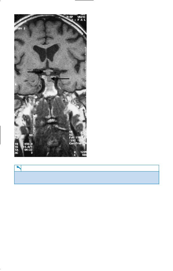
Figure 58.1 Magnetic resonance imaging scan through the pituitary.
KEY POINTS
•The commonest causes of polyuria are diabetes mellitus and chronic renal failure.
•Breast carcinoma may recur after several years of remission.
155
This page intentionally left blank

CASE 59: BLOOD IN THE URINE
History
A 52-year-old businessman is referred to a nephrologist for investigation of microscopic haematuria. This was first detected 6 months ago at an insurance medical for a new job, and has since been confirmed on two occasions by his general practitioner (GP). Previous urinalyses have been normal. He has never had macroscopic haematuria, and has no urinary symptoms. He is otherwise in excellent health. There is no significant past medical history. He has no symptoms of visual problems or deafness. There is no family history of renal disease. He drinks 35 units of alcohol per week and smokes 30 cigarettes per day.
Examination
He is a fit looking well-nourished man. His pulse is 72/min, blood pressure 146/102 mmHg. Otherwise, examination of his cardiovascular, respiratory, abdominal and neurological systems is unremarkable. Funduscopy reveals arteriovenous nipping.
INVESTIGATIONS
|
|
Normal |
Haemoglobin |
13.6 g/dL |
13.3–17.7 g/dL |
Mean corpuscular volume (MCV) |
83 fL |
80–99 fL |
White cell count |
4.2 % 109/L |
3.9–10.6 % 109/L |
Platelets |
213 % 109/L |
150–440 % 109/L |
Sodium |
138 mmol/L |
135–145 mmol/L |
Potassium |
3.8 mmol/L |
3.5–5.0 mmol/L |
Urea |
8.2 mmol/L |
2.5–6.7 mmol/L |
Creatinine |
141 &mol/L |
70–120 &mol/L |
Albumin |
38 g/L |
35–50 g/L |
Glucose |
4.5 mmol/L |
4.0–6.0 mmol/L |
Bilirubin |
13 mmol/L |
3–17 mmol/L |
Alanine transaminase |
33 IU/L |
5–35 IU/L |
Alkaline phosphatase |
72 IU/L |
30–300 IU/L |
Gamma-glutamyl transpeptidase |
211 IU/L |
11–51 IU/L |
Urinalysis: '' protein; '' blood; #100 red cells 24-h urinary protein: 1.2 g; normal !200 mg/24 h Electrocardiogram (ECG): left ventricular hypertrophy Renal ultrasound: two normal-sized kidneys
Questions
•What is the likely diagnosis?
•How do you interpret the creatinine value?
•What further investigations would you organize?
•What advice would you give this patient?
157

ANSWER 59
Microscopic haematuria has many renal and urological causes, e.g. prostatic disease, stones, but the presence of significant proteinuria, hypertension and renal impairment suggest this man has some form of chronic glomerulonephritis. The high gamma-glutamyl transpeptidase level is compatible with liver disease related to a high alcohol intake. The recommended upper limit for men is 28 units per week.
!Commonest glomerular causes of microscopic haematuria
•Immunoglobulin A (IgA) nephropathy
•Thin basement membrane disease
•Alport’s syndrome (predominantly affects males)
IgA nephropathy is the commonest glomerulonephritis in developed countries, and is characterized by diffuse mesangial deposits of IgA. Patients often have episodes of macroscopic haematuria concurrent with upper respiratory tract infection. Most cases of IgA nephropathy are idiopathic, but this it is also commonly associated with Henoch–Schönlein purpura and alcoholic cirrhosis. This man has IgA nephropathy in association with alcoholic liver disease. About 20 per cent of patients with IgA nephropathy will develop end-stage renal failure after 20 years of follow-up.
Thin basement membrane disease is a familial disorder which presents with isolated microscopic haematuria, minimal proteinuria and normal renal function that does not deteriorate. Electron microscopy shows diffuse thinning of the glomerular basement membranes (the width is usually between 150 and 225 nm versus 300–400 nm in normal subjects). Alport’s syndrome is a progressive form of glomerular disease, associated with deafness and ocular abnormalities and is usually inherited as an X-linked dominant condition so that males are more seriously affected.
This patient should have a renal biopsy to reach a histological diagnosis. As the patient is over 50 years old he should have urine cytology/prostate-specific antigen/cystoscopy performed to exclude concurrent bladder and prostatic lesions. He needs a liver ultrasound, and liver biopsy should be considered.
The patient should be advised to abstain from alcohol, and needs his blood pressure controlling. He needs regular follow-up as he is at risk of progressing to dialysis and/or renal transplantation. The raised creatinine appears modest in terms of the actual figures, but as plasma/serum creatinine does not begin to rise until the glomerular filtration rate is reduced to 50 per cent of normal (irrespective of the patient’s age), the raised creatinine in this case indicates a serious loss of renal function to approximately 40 per cent of normal. There is no convincing evidence for immunosuppression retarding the progression into renal failure in most patients with IgA nephropathy.
KEY POINTS
•Patients with isolated haematuria aged !50 years should be initially referred to a nephrologist.
•Patients with isolated haematuria aged #50 years should be initially referred to a urologist for investigation, to exclude bladder or prostatic disease.
•Small elevations in serum/plasma creatinine indicate large loss in renal function.
•Liver damage from a high alcohol intake may occur with no obvious signs and symptoms.
158