Материал: 100_Cases_in_Clinical_Medicine

CASE 93: TIREDNESS
History
A 79-year-old man is brought to his general practitioner by his daughter who says that he is getting very tired and has lost interest in life. She says that a general malaise has been present for 5–6 weeks. She thinks that he might have lost a few kilograms in weight over this time, but he does not weigh himself regularly. He says that he has felt limited on exertion by tiredness for a year or so, and on a few occasions when he tried to do more he had a feeling of tightness across his chest. There is no other medical history of note. He smokes 20 cigarettes a day and drinks a pint or two of Guinness each Saturday and Sunday. He is not on any medication, just taking occasional paracetamol. On systems review, he says that he has lost his appetite over the last month. His sleep has been disturbed by occasional nocturia, and on two or three occasions in the last few weeks he has been disturbed by sweating at night.
There is no relevant family history. He is a retired shopkeeper who normally keeps reasonably fit walking his dog.
Examination
His pulse is 70/min, blood pressure 110/66 mmHg. There is no clubbing, but tar staining is present on the fingers and nails of the right hand. The jugular venous pressure is not raised. The apex beat is displaced 2 cm from the midclavicular line. On auscultation of the heart there is a grade 3/6 ejection systolic murmur radiating to the carotids and a soft early diastolic murmur audible at the lower left sternal edge. There are no abnormalities to find in the abdomen or nervous system. The urine looked clear but routine stick testing showed a trace of blood and on urine microscopy there were some red cells. A chest X-ray was reported as showing a slightly large heart.
INVESTIGATIONS
|
|
Normal |
Haemoglobin |
10.7 g/dL |
13.3–17.7 g/dL |
Mean corpuscular volume (MCV) |
88 fL |
80–99 fL |
White cell count |
12.2 % 109/L |
3.9–10.6 % 109/L |
Neutrophils |
10.5 % 109/L |
1.8–7.7 % 109/L |
Lymphocytes |
1.5 % 109/L |
0.6–4.8 % 109/L |
Platelets |
287 % 109/L |
150–440 % 109/L |
Erythrocyte sedimentation rate (ESR) |
68 mm in 1 h |
!20 mm in 1 h |
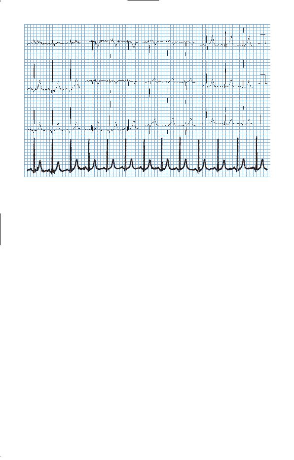
The electrocardiogram (ECG) is shown in Fig. 93.1.
234
I |
aVR |
V1 |
V4 |
II |
aVL |
V2 |
V5 |
III |
aVF |
V3 |
V6 |
II
Figure 93.1 Electrocardiogram.
Questions
•What is the most likely diagnosis?
•What investigations are indicated?
235

ANSWER 93
This 79-year-old man has the clinical features of aortic stenosis and regurgitation. The murmurs are of mixed aortic valve disease and the ECG shows left ventricular hypertrophy (sum of negative deflection in V1 and positive deflection in V5 or V2 and V6 greater than 35 mm), suggesting that there has been significant pressure overload from aortic stenosis. The findings of mixed aortic valve disease, microscopic haematuria, malaise and fever (probable with the night sweats) make infective endocarditis a likely diagnosis. This would fit with the haematological picture showing a normocytic anaemia, a raised neutrophil count and a high ESR. In the elderly, infective endocarditis may be an insidious illness and should be considered in any patient who has murmurs and fever or any other change in the cardiac signs or symptoms. The other classical findings of splenomegaly, splinter haemorrhages, clubbing, Osler’s nodes, Janeway lesions and Roth’s spots are often absent. Precipitating events such as dental treatment or other sources of bacteraemia may not be evident in the history.
It is difficult to tie all the features into any other single diagnosis. The signs are of aortic valve disease. When there is a fever or other evidence of infection in the presence of valve disease, infective endocarditis must always be considered although in practice other unrelated infections are more common. Other infections such as tuberculosis or abscess are possible or an underlying lymphoma or other malignancy.
The most important investigations would be:
•blood cultures performed before any antibiotics are given. In this case three blood cultures grew Streptococcus viridans
•echocardiogram which showed a thickened bicuspid aortic valve, a common congenital abnormality predisposing to significant functional valve disturbance in middle and old age. Vegetations can be detected on a transthoracic echocardiogram if they are prominent, but transoesophageal echocardiogram is more sensitive in detecting vegetations on the valves.
Treatment with intravenous benzylpenicillin and gentamicin for 2 weeks, followed by oral amoxicillin resulted in resolution of the fever with no haemodynamic deterioration or change in the murmurs of mixed aortic valve disease. A microbiologist should be consulted about appropriate antibiotics and duration.
After treatment of the endocarditis, the symptoms of pain and tiredness on exertion would need to be considered to see if valve surgery was indicated. Prior to this it would be routine to look at the coronary arteries by angiography to see if simultaneous coronary artery surgery was needed.
KEY POINTS
•Symptoms on exertion in aortic valve disease are a sign that valve surgery needs to be considered.
•In infective endocarditis, it is unusual to have many of the classical physical signs. In the elderly, it may present with non-specific malaise.
236
CASE 94: ABDOMINAL PAIN
History
A 70-year-old woman is admitted to hospital with acute onset of abdominal pain. The abdominal pain started quite suddenly 24 h before admission and has continued since then. It is a constant central abdominal pain. She has vomited altered food on one occasion.
She has a history of occasional angina on exertion for 5 years. She has a glyceryl trinitrate spray but she has not needed this in the last 3 months. A year ago she was found to be in atrial fibrillation at 120/min, and she was started on digoxin, which she still takes. The only other medical history of note is that she had a hysterectomy for menorrhagia 30 years ago and she has hypertension controlled on a small dose of a thiazide diuretic for the last 3 years. She does not take any other medication apart from low-dose aspirin. She does not smoke and does not drink alcohol. She retired from work as a cleaner 8 years ago.
Examination
She was in atrial fibrillation at a rate of 92/min with a blood pressure of 114/76 mmHg. Respiratory examination was normal. She was tender with some guarding in the centre of the abdomen. No masses were palpable in the abdomen and there were just occasional bowel sounds to hear on auscultation. Over the next 2 h the blood pressure fell to 84/60 mmHg. She was admitted to the intensive care unit (ICU) and monitored while initial investigations were performed. The abdominal X-ray showed no gas under the diaphragm and no dilated loops of bowel or fluid levels. While under observation, the urine output fell off. Re-examination showed that bowel sounds were absent. Her hands and feet remained warm. Measurements of cardiac output in ICU showed that it remained high.
INVESTIGATIONS
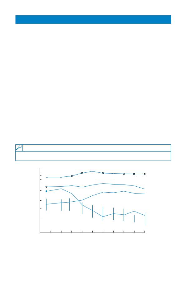
The observation charts are shown in Fig. 94.1.
|
40 |
|
|
|
|
|
39 |
|
|
|
Central |
Temperature |
38 |
|
|
|
temperature |
37 |
|
|
|
|
|
(°C) |
36 |
|
|
|
Peripheral |
|
35 |
|
|
|
|
|
34 |
|
|
|
temperature |
|
|
|
|
|
Pulse |
Blood |
100 |
|
|
|
|
pressure |
|
|
|
|
|
(mmHg) |
8 |
|
|
|
|
|
|
|
|
|
|
CVP |
|
|
|
|
Central venous |
4 |
|
|
|
pressure |
|
(mmHg) |
|
|
|
||
|
|
|
|
||
|
|
|
|
|
|
|
|
|
|
|
Blood pressure |
|
1 |
2 |
3 |
4 |
5 |
|
|
|
|
|
Hours |
Figure 94.1 Chart from intensive care unit.
Questions
•What is the likely cause of the abdominal pain?
•What further developments do the charts suggest?
237

ANSWER 94
One diagnosis of the abdominal pain which would explain her condition and fit with her predisposing situation is ischaemic bowel caused by an embolus from the heart. The patient is likely to become very ill without markedly abnormal physical signs. Atrial fibrillation increases the likelihood of such an event. She has been on aspirin which will reduce slightly the risk of embolic events, but not on anticoagulants which would have decreased the risk further. In the presence of pre-existing cardiovascular problems, shown by the hypertension and angina, anticoagulation would normally be started if there are no contraindications. The risk of cerebrovascular accidents caused by emboli from the heart has been shown to be reduced. In lone atrial fibrillation with no underlying cardiac disease the risks of emboli and the benefits of anticoagulation are less. There are alternative diagnoses such as perforation or pancreatitis, and it is not possible to be sure of the cause of the abdominal problem from the information given here.
The chart of the observations (Fig. 94.1) covers 10 h. After the first hour or two the central venous pressure drops, the blood pressure falls and the pulse rate rises in association with the fall in urine output.
These findings show that she is developing shock with inadequate perfusion of vital organs.
! Possible causes for shock
Types of shock |
Example |
Hypovolaemic shock |
Blood loss |
Cardiogenic shock |
Myocardial infarction |
Extracardiac obstructive shock |
Pulmonary embolism |
Vasodilatory (distributive) shock |
Sepsis |
All these causes are possible in this woman with abdominal problems and a history of ischaemic heart disease. The fact that the cardiac output is high makes blood loss and cardiogenic shock unlikely. The most likely cause is septic shock where peripheral vasodilatation would lead to a high cardiac output but a falling blood pressure and rising pulse rate. Vasoconstriction and reduced blood flow occurs in certain organs, such as the kidneys, leading to the term ‘distributive shock’ with maintained overall cardiac output but inappropriate distribution of blood flow. The rise in central temperature and the lack of a marked fall in peripheral temperature would fit with this cause of the shock.
The patient was stabilized with fluid replacement and antibiotics before going to theatre where the diagnosis of ischaemic bowel from an embolus was confirmed. Arteriography can confirm the diagnosis but confirmation is often at laparotomy which is usually required to remove the necrotic bowel.
KEY POINTS
•Aspirin and anticoagulation should be considered in patients with atrial fibrillation.
•Septic shock may be present with warm peripheries through vasodilatation.
•A drop in the central venous pressure may be the first sign of developing shock.
238