Материал: 100_Cases_in_Clinical_Medicine

CASE 91: PAIN IN THE FOOT
History
A 65-year-old man presents with an ulcer on the dorsum of his right foot. He noticed a sore area on the right foot 3 weeks ago and this has extended to an ulcerated lesion which is not painful. He has complained of pain in the legs for some months. This pain comes on when he walks and settles down when he stops.
He had an inguinal hernia repaired 2 years ago and he stopped smoking then on the advice of the anaesthetist. Previously he smoked 20 cigarettes per day. He drinks four pints of beer at weekends. His father died of a myocardial infarction aged 58 years.
Examination
His blood pressure is 136/84 mmHg. The respiratory, cardiovascular and abdominal systems are normal. There is a 3 cm ulcerated area with a well-demarcated edge on the dorsum of the right foot. The posterior tibial pulses are palpable on both feet, and the dorsalis pedis on the left. The capillary return time is 4 s. On neurological examination there is some loss of light touch sensation in the toes. Varicose veins are present in the long saphenous distribution on both legs.
INVESTIGATIONS
|
|
Normal |
Haemoglobin |
14.3 g/dL |
13.7–17.7 g/dL |
White cell count |
7.4 % 109/L |
3.9–10.6 % 109/L |
Neutrophils |
4.6 % 109/L |
1.8–7.7 % 109/L |
Lymphocytes |
2.5 % 109/L |
0.6–4.8 % 109/L |
Platelets |
372 % 109/L |
150–440 % 109/L |
Sodium |
140 mmol/L |
135–145 mmol/L |
Potassium |
4.0 mmol/L |
3.5–5.0 mmol/L |
Urea |
5.1 mmol/L |
2.5–6.7 mmol/L |
Creatinine |
89 &mol/L |
70–120 &mol/L |
Glucose |
6.4 mmol/L |
4.0–6.0 mmol/L |
HbA1c |
9.1 per cent |
!7 per cent |
Question
• What is the likely diagnosis?
229

ANSWER 91
The presence of varicose veins raises the possibility of a venous ulcer related to poor venous return. However, venous ulcers are usually found around the medial malleolus and are often associated with skin changes of chronic venous insufficiency. This has the features of an ulcer caused by arterial rather than venous ulceration or a mixed aetiology. Arterial ulcers are often on the dorsum of the foot. Arterial ulcers tend to be deeper and more punched out in appearance. The left dorsalis pedis pulse is not palpable and the capillary return time is greater than the normal value of 2 s. The story of pain in the legs on walking requires a little more detail but it is suggestive of intermittent claudication related to insufficient blood supply to the exercising calf muscles.
The raised HbA1c suggests diabetes and prolonged hyperglycaemia. In diabetes the arterial involvement may be in small vessels with greater preservation of the pulses. The peripheral sensory neuropathy may also be associated with diabetes and lead to unrecognized trauma to the skin which then heals poorly. Other risk factors for arterial disease are the family history and the history of smoking.
Further investigations would include measurement of the ankle:brachial blood pressure ratio. If this is less than 0.97 it suggests arterial disease, and a low index would be a contraindication to pressure treatment in trying to heal the ulcer.
Ultrasonic angiology would help to identify the anatomy of the arterial circulation in the lower limbs and would show if there are correctable narrowings of major vessels. Good control of diabetes can slow progression of complications such as neuropathy and microvascular disease. Care of the feet is a very important part of the treatment of diabetes and should be a regular element of follow-up.
KEY POINTS
•The position and nature of ulcers provide clues to their cause.
•Diabetic feet are particularly vulnerable because of sensory loss, arterial insufficiency and high sugars. Foot care is an important element of regular diabetic management.
230
CASE 92: A HEALTHY MAN?
History
A 50-year-old man has a health screen as part of an application for life insurance. He has no symptoms. He smokes 15 cigarettes per day and drinks 10 units of alcohol per week. In his family history his father died of a myocardial infarction aged 56 years.
Examination
He weighs 84 kg and is 1.6 m (5 ft 8 in) tall. His blood pressure is 164/98 mmHg. Examination is otherwise normal.
INVESTIGATIONS
|
|
Normal |
Haemoglobin |
15.2 g/dL |
13.3–17.7 g/dL |
White cell count |
10.0. % 109/L |
3.9–10.6 % 109/L |
Platelets |
287 % 109/L |
150–440 % 109/L |
Sodium |
139 mmol/L |
135–145 mmol/L |
Potassium |
3.9 mmol/L |
3.5–5.0 mmol/L |
Urea |
4.3 mmol/L |
2.5–6.7 mmol/L |
Creatinine |
88 &mol/L |
70–120 &mol/L |
Cholesterol |
5.0 mmol/L |
!5.5 mmol/L |
Triglyceride |
1.30 mmol/L |
0.55–1.90 mmol/L |
Very low-density lipoprotin (VLDL) |
0.44 mmol/L |
0.12–0.65 mmol/L |
Low-density lipoprotein (LDL) |
3.1 mmol/L |
1.6–4.4 mmol/L |
High-density lipoprotein (HDL) |
1.9 mmol/L |
0.9–1.9 mmol/L |
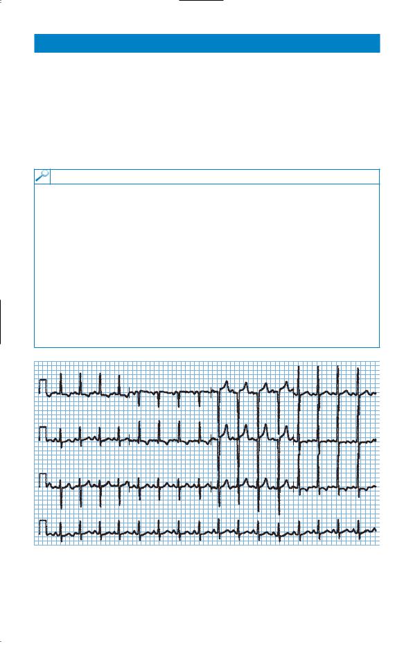
His electrocardiogram (ECG) is shown in Fig. 92.1.
I |
aVR |
V1 |
V4 |
|
II |
aVL |
V2 |
V5 |
|
|||
III |
aVF |
V3 |
V6 |
|
II
Figure 92.1 Electrocardiogram.
Question
• What is the appropriate management?
231

ANSWER 92
The ECG shows left ventricular hypertrophy (R-wave in V5 and S-wave in V1 #35 mm). Although only a single reading is given, the hypertrophy makes it likely that the blood pressure represents sustained hypertension rather than a ‘white coat’ effect. It should be repeated several times over the next few weeks for confirmation, but treatment is likely to be indicated.
The risks of vascular disease are related to the presence of other risk factors. The body mass index is 28 showing that he is overweight. He is a smoker with a positive family history of cardiovascular disease. Tables such as the Sheffield table can be used to obtain a calculation of the risks of cardiovascular disease.
The other question is whether a search for the cause of the hypertension is indicated. Around 85 per cent of cases are idiopathic. Most of the secondary cases are related to renal disease, and the renal function is normal here. A number of endocrine causes (Cushing’s syndrome, Conn’s syndrome) are associated with hypokalaemia. If the blood pressure is difficult to control, secondary causes such as renal artery stenosis should be considered and investigated by renal ultrasound or a technique to visualize the renal arteries such as magnetic resonance angiography or digital subtraction angiography.
The cholesterol is at a level which would warrant treatment if there was evidence of vascular disease. The hypertension itself should be controlled according to current guidelines which would recommend starting with an angiotensin-converting enzyme (ACE) inhibitor in a patient younger than 55 years.
KEY POINTS
•A single elevated blood pressure needs to be remeasured over several weeks.
•All relevant risk factors should be considered in assessing cardiovascular risk and planning treatment.
•Most cases of hypertension do not have an identifiable underlying cause.
232
This page intentionally left blank