Материал: BASIC_SURGICAL_TECHNIQUES budapest
4.1.3. Instruments used for hemostasis
They act mechanically or thermally to stop bleeding at the site of incision or in the surgical territory. The important members of this group are: vascular clamps (Péan, mosquito, abdominal Péan, Kocher, Lumnitzer, Satinsky, bulldog), electrocautery knife, various ligation needels and directing probes (e.g. Deschamp ligation needle, and Payr probe), and argon beam coagulator.
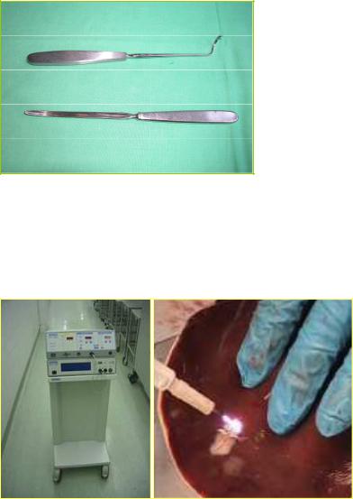
Deschamp ligation needle and Payr sonda (probe)
The Payer probe is used to dissect the area which is located beneath the vessel. Following this, it is kept under the vessel and the Deschamp ligation needle is directed under the vessel and above the probe. Suture material is passed through the hole found at the end of the Deschamp needle which is then directed back from under the vessel. In this manner, we can ligate the desired vessel (Figure 30.).
A
B
Figure 30. Deschamp ligation needle (A) and Payr probe (B)
Argon beam coagulator
It is one of the newest instruments for hemostasis during the operations performed on solid organs. It makes possible to do a monopolar coagulation with a so-called ”no-touch technique”. Its penetration depth is small, for this reason the hemostasis is rapid and effective (Figure 31.).
Figure 31. Argon beam coagulator
31
4.1.4. Reratcting instruments
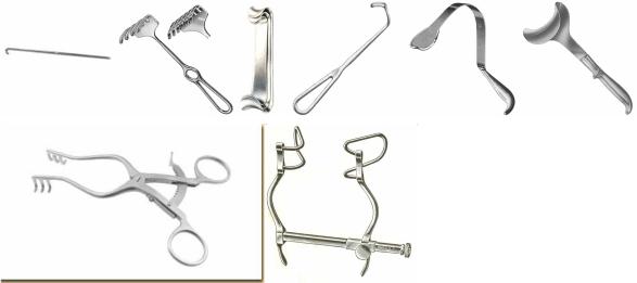
Retarctors are used to hold tissues and organs aside in order to improve the exposure and hence the visibility and accessibility of the surgical field. Hand-held retractors (e.g. skin hook, rake, Roux, Langenbeck, visceral and abdominal wall retarctors) are held by assistant. They cause minimal tissue damage because the assistant maintains tension on tissues only as long as necessary. When applied properly, self-retaining retractors (e.g.Weitlaner self-retaining retractor, Gosset self-retaining retractor) are of great help, but care should be taken not to damage the tissues when they are placed and removed (Figure 32.).
A |
B |
C |
D |
E |
F |
|
|
|
|
|
|
G |
|
H |
|
|
|
Figure 32. Reratcting instruments
A. Skin hook, B. Rake retractor, C. Roux retractor, D. Langenbeck retractor, E. Visceral retractor, F. Abdominal wall retractor, G. Weitlaner self-retaining retractor, H. Gosset selfretaining retractor
4.1.5. Wound-closing instruments and materials
The instruments (and materials) used to unite the tisses belong to this group. The basic principle for wound healing is the proper and tension-free approximation of tissues. Next to this, any dead space should be avoided, as well as there should be an appropriate blood supply of the tissues. The number of stiches (or clips) should be as little as needed. The surgical needles, suture materials, needle holder (see ”grasping instruments”), staplers, clips, and adhesive tapes belong to this group.
Surgical needles and sutures
Detailed disscution of this part can be found in section 4.2.
Staplers
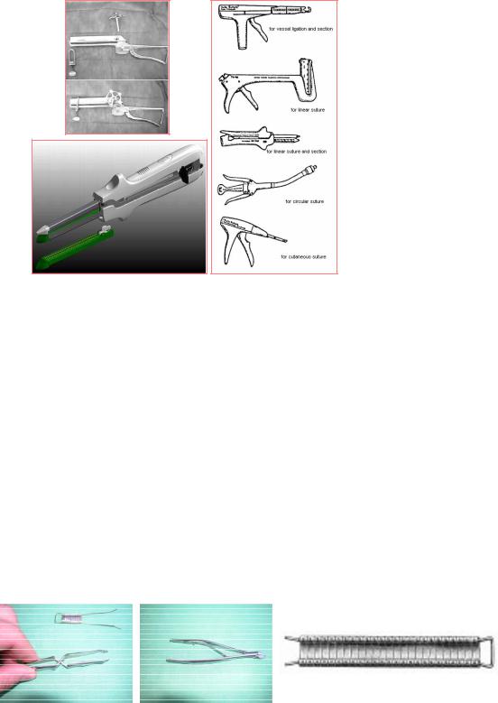
Suturing of a big surgical area is exhausting. Beside this, the pulling of the tissue can be a cause for a later insufficiency and the operation time is also increased in the case of the hand suturing. Due to these reasons and especially in intestinal or lung surgeries, the staplers are essentially important. The staplers can be either linear-which produce the suture row along a straight line -or circularwhich are used to make anastomosis between two hollow organs (Figure 33.).
32
A C
B
Figure 33. Staplers
A. Petz stapler, B. Linear stapler, C. Staplers with various shapes for different uses
Clips
The classic Michel clips -which can be used with the help of Michel clip applicator and remover-, are used to close a skin wound (Figure 34.). Clips are generally useful for closure of any luminal structure (e.g. vessel, duct).
Other uses of metalic clip:
-in the wound stapler, which makes possible the atraumatic and fast closure of the wound
-in hemostasis (The metalic clips can occlude the lumen of the vessel well)
-the metalic clip can be seen in x-ray film. So it can be useful for various markings (e.g., the bed of a tumor)
Appearances of the CT and MRI have changed our views relating to the use of the
metallic clips. In the case of CT, the clip disturbs the picture only in the vicinity of it and so the examination can be done. In the csae of MRI, the implanted or used metal (e.g. iron, nickle, cobalt) makes impossible to perform the examiation because these metals can move in a magnetic field and in this way a vascular clip can fall down or the intracranial clips can become wandering. Due to spreading of the MRI examinations, it is advisable to use the non-magnetic clips (e.g. titanium, platinum, and absorbable clips).
|
|
|
|
|
|
|
|
|
|
A |
|
B |
|
C |
|
|
|
|
|
Figure 34. Michel clip applicator (A), Michel clip remover (B), Michel clips (C)
33
Self-adhesive strips
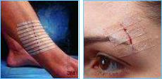
Self-adhesive strips (Stri-Strip) can also unify the tissues. They can be applied in the case of smaller wounds not requiring suturing, when the wound edges can easily and well be approximated. They are also used to fasten the subcuticular sutures. Their use is easy and fast but they can only be used in dry and non-secreting areas (Figure 35.).
Figure 35. Self-adhesive strips
Surgical adhesives
They are usually produced from fibrin, collagen or thrombin and induce the last phase of blood coagulation, so that a firm fibrin mesh is produced. Application fields: for hemostasis in operations done on solid organs, and to close the place of air leakage in lung surgeries. Disadvantages: in infected wounds, they can increase the degree of infection and lead to abscess formation.
4.1.6. Special instruments
Those instruments which are not used routinely during surgical interventions belong to this group.
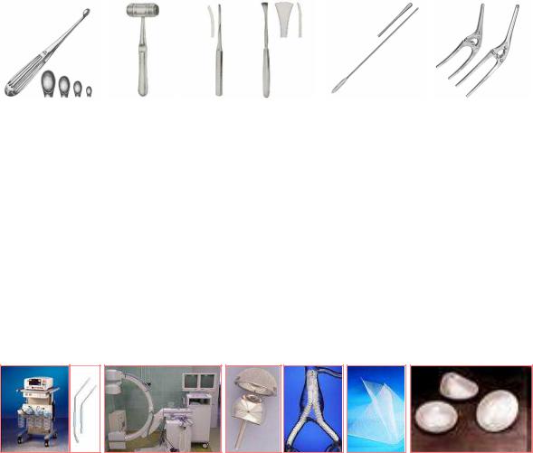
Volkmann curette
They have various sizes. The egdes of the distal spoon-shaped part of this instrument are sharp which make possible to remove the tissues. The main application areas: skin tags (e.g. condyloma, wart) removal, to clean the base of the infected wound, and to remove the infected bone in the case of osteomyelitis (Figure 36. A).
Instruments used in bone surgeries
They are helpful to perform operations on the bones in orthopaedic surgery and traumatology (Figure 36. B and C).
Round-ended probe
They are straight or curved malleable metalic rods with various sizes. Their end is generally rounded. Use to gauge depth or direction of a sinus or cavity by inserting it there in (Figure 36. D).
Payr clamp
We use it before resecting of the intestine. The essence of the crushing is to apply equal pressure over the serosal layer whereby we can avoid the tearing of the serosa before application of the ligature (Figure 36. E).
34
A |
|
B |
C |
|
|
|
D |
|
E |
|
|
|
|
|
|
|
|
|
|
Figure 36. Special instruments
A. Volkmann curette, B. Mallet, C. Chisels, D. Round-ended probe, E. Payr clamp
Suction set
It is used to suck the larger amounts of the blood and secretions from the surgical territories. This set consists of a resterilizable suction tip, a tube and a nonsterile container. The uper end of the conatiner is connected to a central suction system.
X-raying set
It is mainly used during the operations done on bones.It makes possible to check the position of the bones and the implanted metals during the surgery.
Implants, protheses
The metalic screws and pins, joint protheses, hernial meshes, vascular grafts and silicon implants (used in breast surgery) belong to this group.
A B C D E F
Figure 37. Special instruments
A. Suction set and suction tips, B. X-raying set, C. Metalic joint prothesis, D. Vascular grafts, E. Hernial meshes, F. Breast implants
4.2. Suturing tools and materials
4.2.1. Surgical needles
In the history of healing, many materials were used as the surgical needels (e.g. bone, fish bone, and acacia thorn). Since 19th century the metalic needle-which was non-disposable for a long period of time-has been used.
The criteria for an ideal needle:
-should be made of the stainless steel with a high quality, which causes minimal tissue reaction,
-should be thin as much as possible (but this should not affect the strength of the needle),
-can be fixed and directed in a stable manner on the needle holder,
-can direct the suture material with a suitable assurance and a minimum tissue injury,
-should be sharp enough to pass through the tissues (with a minimum tissue resistance)
-should be stiff enough to resist bending but at the same time it shloud be flexible enough not to
35