Материал: BASIC_SURGICAL_TECHNIQUES budapest
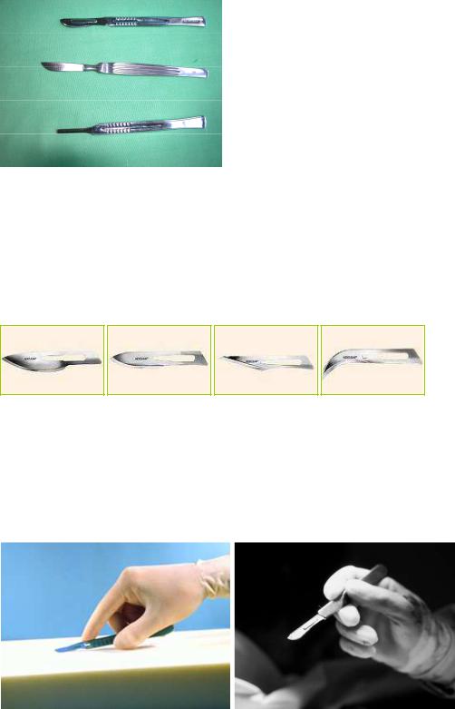
Figure 8. Type of scalpels
A. Handle with a disposable blade, B. Conventional scalpel, C. Handle
Wide-bladed scalpels with a curved cutting edge are used for incising skin and subcutaneous tissues. At all times, the skin incision should be done with scalpel because this will insure the proper adjusting of the edges which is a fundamenal factor in healing process. Cutting with the whole length of the cutting edge (and not merely with its tip) can lead to less injury to the tissue. Thin-bladed, sharp-tipped scalpels serve for the opening of blood vessels, ducts, and abscesses (Figure 9.).
Figure 9. Blades with various sizes and shapes
Holding of the scalpel:
1.In long, straight incisions, the scalpel is held like a fiddle bow: the handle is gripped horizontally between the thumb and middle fingers while the index finger is staying above the handle. The ring and little fingers are holding the end of the handle.
2.In short or fine incisions, the scalpel is held like a pencil, and the cutting is made mostly with the tip (Figure 10.).
A |
|
B |
|
|
|
|
|
|
Figure 10. Holding of the scalpel
A. Fiddle bow holding, B. Pencil holding
21
Scissors
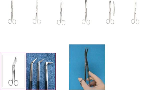
Next to the scalpel, scissors are most often used to dissect and cut tissues. Threads and bandages are also cut with scissors. Scissors can be of different sizes. Their blade can be straight, curved or angular. The tips of the blades can be blunt-blunt, blunt-sharp or sharp-sharp. The cutting is ususally made by portion of the blade which is close to its tip (Figure 11. and 12.). Scissors are also suitable for blunt dissection and preparation of the tissues. In this case the scissors are introduced into the tissues with their tips closed. Thereafter, we open the scissors and do the dissection with the lateral blunt edges of the blades.
A |
|
B |
|
C |
|
D |
|
E |
|
F |
|
|
|
|
|
|
|
|
|
|
|
Figure 11. Scissors with various tips and blades: A. Straight blunt-blunt scissors, B. Straight blunt-sharp scissors, C. Straight sharp-sharp scissors, D. Curved blunt-blunt scissors, E. Curved blunt-sharp scissors, F. Curved sharp-sharp scissors
AB
Figure 12. Scissors which are angled at the joints: A. Lister bandage scissors, B. Kneed scissors
Figure 13. Correct holding of the scissors (1st-4th rule of holding the instrument)
Correct holding of the scissors and all ring-ended instruments: the thumb and the fourth finger are inserted into the rings which are located at the handle, while the index finger is placed distally over the handle to stabilize the scissors (1st-4th rule of holding the instrument) (Figure 13. and 14.).
22
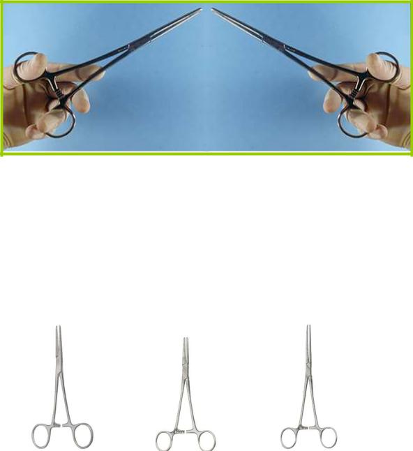
Figure 14. Correct holding of the ring-ended instruments with right and left hands
Hemostats used for tissue preparation: Péan clamp, mosquito clamp, abdominal Péan clamp
Instruments listed here are suitable for tissue preparation (dissecting instruments), grasping (grasping instruments), as well as haemostasis. They are used for blunt dissection and preparation of the tissues. In this case they are introduced into the tissues with their tips closed. Thereafter, we open them and do the dissection with the lateral blunt edges of the instrument.
Structurally, they are similar to the scissors. There are rings at the proximal end of the handle. A little bit below the rings you can find the locks, which are used to close the handle. Péan clamp, mosquito clamp, and abdominal Péan clamp are traumatic (crushing) clamps (or forceps) because their grasping parts are serrated (Figure 15.).
A |
B |
C |
|
|
|
Figure 15. Hemostats used for preparation
A. Péan clamp, B. Mosquito clamp, C. Abdominal Péan clamp
These instruments can stop bleeding when applied after the preparation of the vessel and before its cutting (planned hemostasis) or used to grasp and clamp the end of a cut vessel which is bleeding. These are ring-ended instruments. So the 1st-4th rule of holding the instruments is applied here. The lock can be opened by pressing down one of the finger rings with our thumb while elvating the other one with the ring finger. In this manner the interlocking teeth are moved from one another. We should learn how to use such these instruments with both of our hands. At the time of their removal we should avoid their twitching and handle them carefully to avoid the
23
tearing of the tissues.
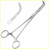
Dissector
Long-handled, ring-ended instrument, which is bended 90° at its distal part. It may or may not have the interlocking teeth. We use them to dissect and prepare the tissues atraumatically (Figure 16.)
Figure 16. Dissector
Diathermy knife
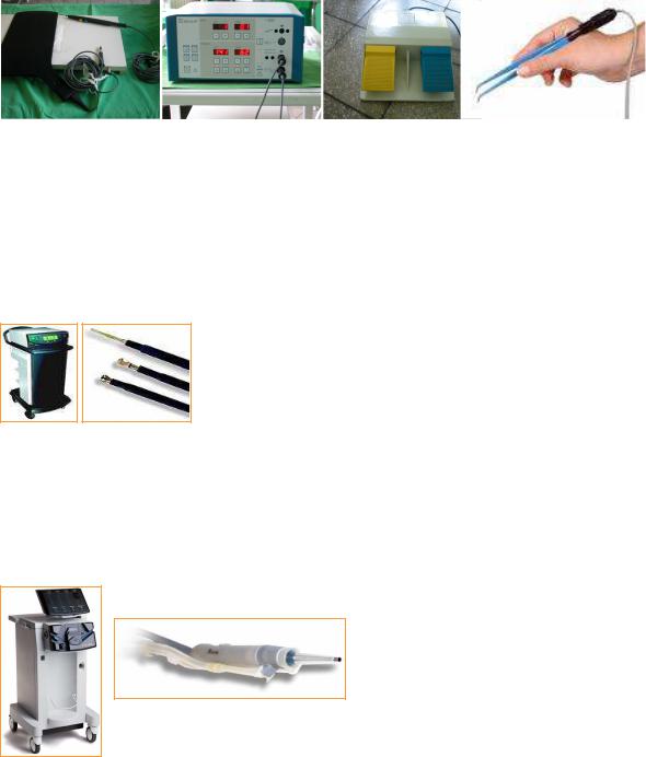
It dissects the tissues with the help of the heat which is generated by the electric current.Its advantage is that during the dissection the heat can also coagulate the small vessels and in this way cutting and hemostasis are happening simultaneously. The diathermy can be either monoor bi-polar. When the electric current is passing between the two parts of the instrument we call it the bipolar diathermy (e.g. bipolar forceps) and when it passes between the instrument and the indifferent electrode -which is placed beneath the back or one of the lower limbs of the patientit is called the monopolar diathermy (e.g. electrocauter or electrocautery knife).
In general surgery the monopolar diathermy is used most commonly. Considering the fact that during the dissection it also coagulates the small vessels, the preparation phase of the the operation will become easier and shorter. In a patient with a pacemaker, the electric current of diathermy can cause arrhythmia. The old type of pacemaker needs to be adjusted prior to the surgery, while with the modern pacemakers this problem does not exsist. It is not advisable to use the diathermy for making a skin incision because it can burn the skin and lead to its necrosis. You should be careful when using it during the operation and for purposes other than skin incision. Because the electric current and heat can be conducted to the skin by any metalic instrument and this itself may again be a cause for the skin necrosis. With use of various voltage and amperage you can only coagulate (the so-called ”coagulation grade” which can be achieved by pressing the blue bottom of the electrocautery). With increasing the voltage and amperage of the device it can become suitable for tissue dissection as well (the so-called ”cutting grade” which can be achieved by pressing the yellow bottom of the electrocautry). These two types of function can also be achieved with use of a foot pedal.In this way other metalic instruments (e.g. forceps) can be used which can lead to a more precise and faster operation (Figure 17. A, B, and C).
In the case of a bipolar diathermy there is a need for smaller voltage and amperage. It makes possible to perform a more precise work and the size of the burned area is smaller as well. The wire of the bipolar forceps is connected to the diathermy device (Figure 17. B and D).
24
A |
B |
C |
D |
|
|
|
|
|
|
Figure 17. Monopolar and bipolar diathermies
A. Monopolar diathery with its indifferent electrode and hand portion, B. Diathermy device (for both monoand bi-polar diathermies, C. Foot pedal of the monopolar diathermy, D. Bipolar forceps
Ultrasonic cutting device
Ultrasonic cutting device (Ultracision®) is using the ultrasound to cut and coagulate the tissues. It is working similarly to the diathermy but the ultrasonic device does not cause a thermic injury. It makes possible to have more precise movements during operation (Figure 18.).
Figure 18. Ultrasonic cutting device and various shapes of its hand portion
CUSA (Cavitron Ultrasonic Surgical Aspirator)
The ultrasonic vibrating knife selectively crushes and sucks the tissues which contain high quantity of water and low amount of collagen; meanwhile it is taking care of other tissues (e.g. vessels and nerves). During operating on solid organs the use of this instrument leads to less blood loss and tissue damage (no thermic injury!), as well as better viewing (Figure 19.).
Figure 19. CUSA and its hand portion
LASER (Light Amplification by Stimulated Emission of Radiation)
CO2 laser is useful for superficial treatment, while the neodymium-YAG-laser is good for 3-5 mm deep areas. Use: cutting, coagulation, vaporization, selective obliteration of the diseased tissues, and palliative treatment of the nonresectable gastrointestinal tumors.
25